. 2007 Feb;143(2):924-40.
doi: 10.1104/pp.106.087668. Epub 2006 Dec 22.

Transcriptional profiling of the Arabidopsis embryo

Affiliations

Transcriptional profiling of the Arabidopsis embryo

Matthew W B Spencer et al. Plant Physiol. 2007 Feb.

Erratum in

  • Plant Physiol. 2007 Apr;143(4):1982

Abstract

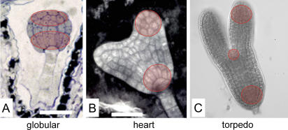
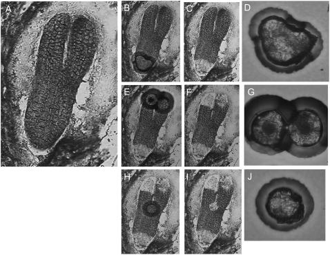
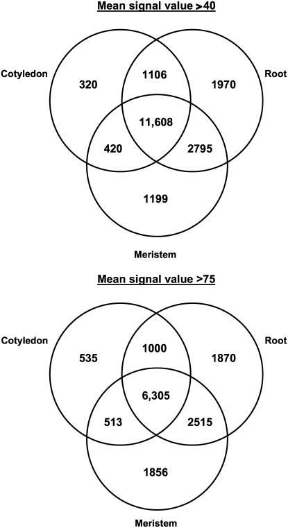
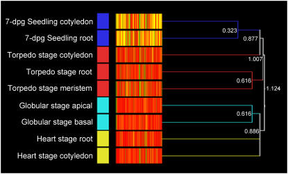
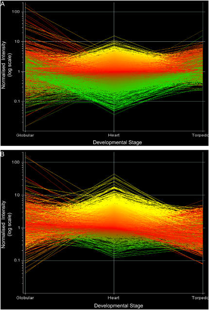
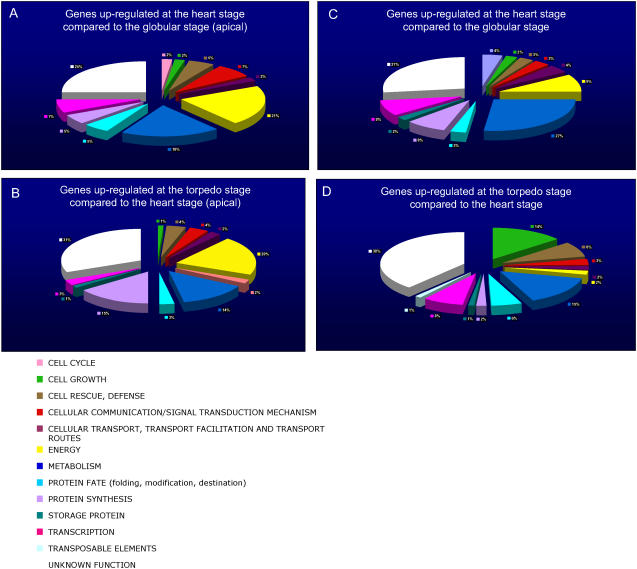
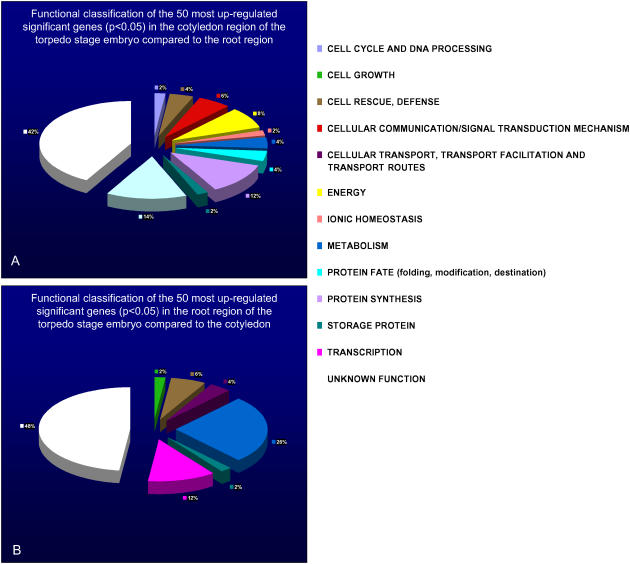
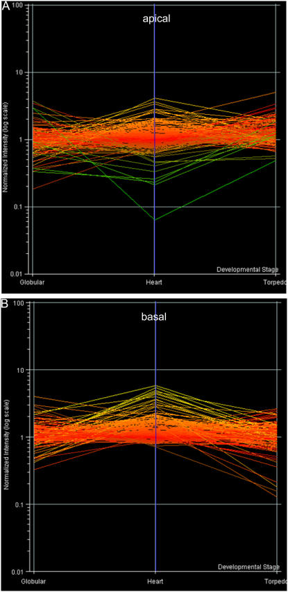
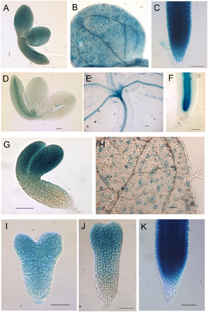
We have used laser-capture microdissection to isolate RNA from discrete tissues of globular, heart, and torpedo stage embryos of Arabidopsis (Arabidopsis thaliana). This was amplified and analyzed by DNA microarray using the Affymetrix ATH1 GeneChip, representing approximately 22,800 Arabidopsis genes. Cluster analysis showed that spatial differences in gene expression were less significant than temporal differences. Time course analysis reveals the dynamics and complexity of gene expression in both apical and basal domains of the developing embryo, with several classes of synexpressed genes identifiable. The transition from globular to heart stage is associated in particular with an up-regulation of genes involved in cell cycle control, transcriptional regulation, and energetics and metabolism. The transition from heart to torpedo stage is associated with a repression of cell cycle genes and an up-regulation of genes encoding storage proteins, and pathways of cell growth, energy, and metabolism. The torpedo stage embryo shows strong functional differentiation in the root and cotyledon, as inferred from the classes of genes expressed in these tissues. The time course of expression of the essential EMBRYO-DEFECTIVE genes shows that most are expressed at unchanging levels across all stages of embryogenesis. We show how identified genes can be used to generate cell type-specific markers and promoter activities for future application in cell biology.


Figures

Figure 1.
Figure 1.
LCM of embryonic tissues. Tissues captured by LCM at globular (A), heart (B), and torpedo (C) stages of embryogenesis, as indicated by highlighted areas.
Figure 2.
Figure 2.
LCM of cryosections of torpedo stage Arabidopsis embryos. A, Torpedo stage embryo. B, Basal region after targeting with laser, after removal of the cap (C), and basal cells captured on cap (D). E, Targeting of the cotyledonary region, after removal of the cap (F), and cotyledonary cells on the cap (G). H, Targeting of the SAM region, after removal of the cap (I), and SAM cells on the cap (J).
Figure 3.
Figure 3.
Venn diagrams showing overlapping expression of genes between the tissue regions of the torpedo stage embryo. A, Venn diagram is presented for signal cutoff values of 40 and 75, with the overlapping regions corresponding to the number of expressed genes present in more than one tissue type. The central region corresponds to the expressed genes present in all tissue types.
Figure 4.
Figure 4.
Cluster analysis of the entire transcriptional profile of roots (basal) and cotyledons (apical) of the developmental stages of globular, heart, torpedo, and seedling and torpedo stage SAM. All samples were initially normalized together to a per-gene median value. Clustering analysis was then performed using condition tree clustering on all samples. The branch values are calculated as distance, i.e. dissimilarity of expression between data clusters; low values represent relatively low dissimilarity, or relatively high similarity (GeneSpring version 7.2).
Figure 5.
Figure 5.
Transcriptional changes across a developmental time course (globular, heart, and torpedo stage embryos) in apical (A) and basal (B) domains. All samples were normalized together to a per-gene median value, indicated in red. Genes expressed to levels higher than the median values are indicated in yellow, while genes expressed to lower levels are indicated in green. All genes on the GeneChip are represented.
Figure 6.
Figure 6.
Functional annotations of the 100 most up-regulated genes (passing the significance filter: P < 0.05 in at least one stage) between developmental stages on the apical developmental (A and B) and basal (C and D) time course, respectively. The color keys show the designated gene class annotations.
Figure 7.
Figure 7.
Functional annotation of the 50 most differentially expressed (by fold-change) significant genes (P < 0.05) between the cotyledon and root regions of the torpedo stage embryo, respectively. A, Genes relatively highly expressed in the cotyledon. B, Genes relatively highly expressed in the root. The color key shows the designated gene class annotations.
Figure 8.
Figure 8.
Transcriptional changes of 222 EMB genes across a developmental time course (globular, heart, and torpedo stage embryos) in apical (A) and basal (B) domains. All samples were normalized together to a per-gene median value, indicated in red. Genes expressed to levels higher than the median values are indicated in yellow, while genes expressed to lower levels are indicated in green.
Figure 9.
Figure 9.
Promoter-GUS fusion activities for genes identified as either constitutively expressed in embryos (A–C) or preferentially expressed in either basal (D–F), apical (G and H) domains, or changing pattern during development (I–K). A to C, At5g45600. GUS activity in cotyledonary stage embryo (A), cotyledon at 3 dpg (B), and primary root tip at 3 dpg (C). D to F, At2g31510. GUS activity in cotyledonary stage embryo (D), hypocotyl and young shoot at 3 dpg (E), and primary root tip at 3 dpg (F). G and H, At5g14610. GUS activity in cotyledonary stage embryo (G) and leaf stomatal guard cells (H). I to K, At5g50810. GUS activity in heart stage (I) and torpedo stage (J) embryo, and primary root tip at 3 dpg (K).

References

    1. Alonso JM, Stepanova AN, Leisse TJ, Kim CJ, Chen H, Shinn P, Stevenson DK, Zimmerman J, Barajas P, Cheuk R, et al (2003) Genome-wide insertional mutagenesis of Arabidopsis thaliana. Science 301 653–657 - PubMed
    1. Ausubel F, Benfey P (2002) Arabidopsis functional genomics. Plant Physiol 129 393. - PMC - PubMed
    1. Barton KM, Poethig RS (1993) Formation of the shoot apical meristem in Arabidopsis thaliana: an analysis of development in the wild type and in the shoot meristemless mutant. Development 119 823–831
    1. Beemster GTS, De Veylder L, Vercruysse S, West G, Rombaut D, Van Hummelen P, Galichet A, Gruissem W, Inzé D, Vuylsteke M (2005) Genome-wide analysis of gene expression profiles associated with cell cycle transitions in growing organs of Arabidopsis. Plant Physiol 132 734–743 - PMC - PubMed
    1. Benjamini Y, Hochberg Y (1995) Controlling the false discovery rate: a practical and powerful approach to multiple testing. J R Stat Soc Ser B 57 289–300

Publication types

MeSH terms

LinkOut - more resources