. 2007 Mar;189(5):1756-64.
doi: 10.1128/JB.01501-06. Epub 2006 Dec 22.

On torque and tumbling in swimming Escherichia coli

Affiliations

On torque and tumbling in swimming Escherichia coli

Nicholas C Darnton et al. J Bacteriol. 2007 Mar.

Abstract

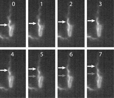
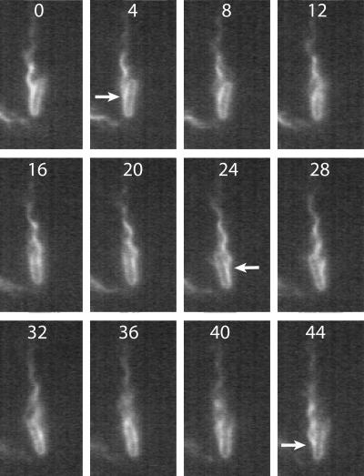
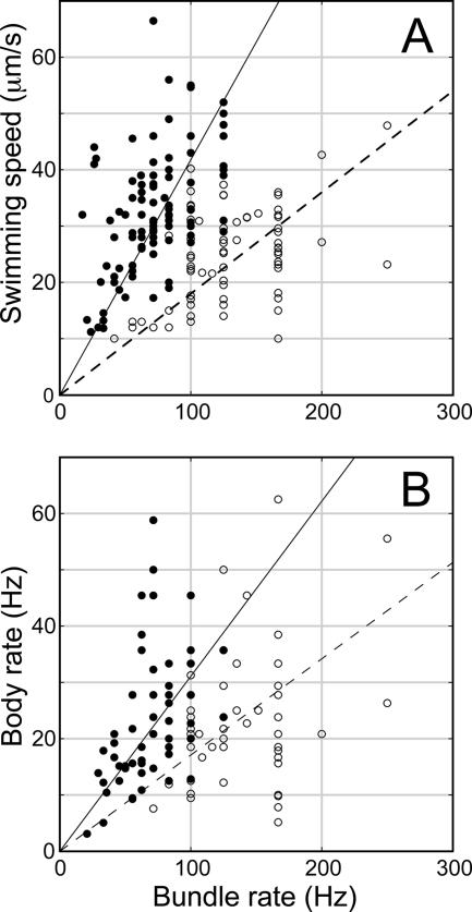
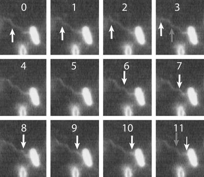
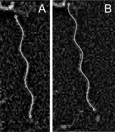
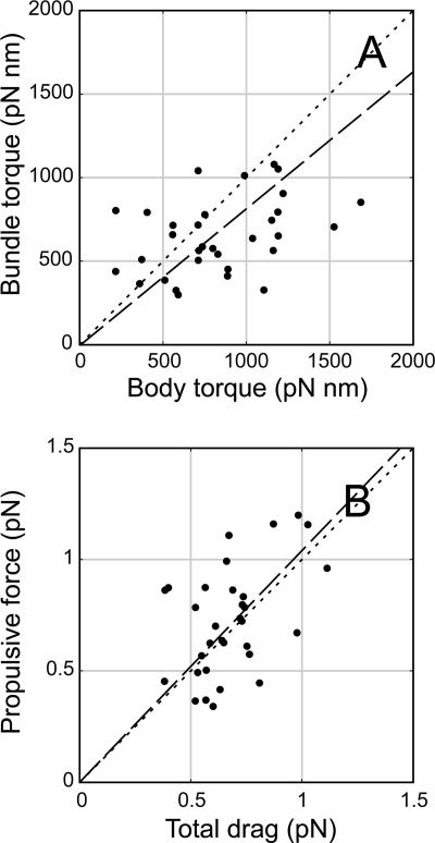
Bacteria swim by rotating long thin helical filaments, each driven at its base by a reversible rotary motor. When the motors of peritrichous cells turn counterclockwise (CCW), their filaments form bundles that drive the cells forward. We imaged fluorescently labeled cells of Escherichia coli with a high-speed charge-coupled-device camera (500 frames/s) and measured swimming speeds, rotation rates of cell bodies, and rotation rates of flagellar bundles. Using cells stuck to glass, we studied individual filaments, stopping their rotation by exposing the cells to high-intensity light. From these measurements we calculated approximate values for bundle torque and thrust and body torque and drag, and we estimated the filament stiffness. For both immobilized and swimming cells, the motor torque, as estimated using resistive force theory, was significantly lower than the motor torque reported previously. Also, a bundle of several flagella produced little more torque than a single flagellum produced. Motors driving individual filaments frequently changed directions of rotation. Usually, but not always, this led to a change in the handedness of the filament, which went through a sequence of polymorphic transformations, from normal to semicoiled to curly 1 and then, when the motor again spun CCW, back to normal. Motor reversals were necessary, although not always sufficient, to cause changes in filament chirality. Polymorphic transformations among helices having the same handedness occurred without changes in the sign of the applied torque.


Figures

FIG. 1.
FIG. 1.
Consecutive images (500 video frames/s) of a cell swimming toward the bottom of the field, propelled by a normal flagellar bundle. The position of an individual helical wavecrest is indicated by white arrows. As the wave propagates away from the cell body, a second crest (gray arrow in frame 5) appears at the original position of the first crest, identifying a complete CCW revolution of the filament. Frame numbers can be converted to elapsed time by multiplying by 0.002 s. Details of this motion are seen more clearly in the movie file “500 Hz swimming.avi” in the supplemental material.
FIG. 2.
FIG. 2.
Every fourth image for the cell shown in Fig. 1. The arrow in frame 4 indicates where a filament arises from the bacterium's surface and joins the bundle. After one-half revolution of the cell body, the bundle appears on the opposite side of the cell (frame 24); after one full revolution, it reappears on the original side of the cell (frame 44). Details of this motion are seen more clearly in the movie file “500 Hz swimming.avi” in the supplemental material.
FIG. 3.
FIG. 3.
Swimming speed (A) and body rotation rate (B) as a function of the bundle rotation rate in MB+ (○) or MB+ with 0.18% methylcellulose (•). The slopes of the linear regression lines are as follows: 0.180 μm for the dashed line and 0.418 μm for the solid line in panel A; and 0.171 for the dashed line and 0.311 for the solid line in panel B.
FIG. 4.
FIG. 4.
Consecutive images (500 video frames/s) of a stuck cell spinning a single flagellar filament. The position of an individual helical wavecrest is indicated by white arrows. As the wave propagates away from the cell body, a second crest (gray arrow in frame 3) appears at the original position of the first crest, identifying a complete CCW revolution of the filament. Frames 4 and 5 are identical; the filament has stopped rotating. The white arrows in frames 6 to 11 indicate the retrograde motion of a helical wavecrest toward the cell body. As the wave propagates toward the cell body, a second crest (gray arrow in frame 11) appears at the original position of the first crest, identifying a complete CW revolution of the filament. Details of this motion are seen more clearly in the movie file “500 Hz reversal 1.avi” in the supplemental material.
FIG. 5.
FIG. 5.
Consecutive images (500 video frames/s) of a stuck cell spinning a single flagellar filament. The position of an individual helical wavecrest is indicated by white arrows as the wave propagates away from the cell body (frames 0 to 4). In frames 5 to 9 filament rotation stops. In frames 10 to 14, the distal end of the filament remains stopped, while a short-pitch region of the transformed filament, indicated by a gray arrow, appears in frame 14. The proximal region is now inclined toward the left of the cell's longitudinal axis (compare frames 1 and 14). Details of this motion are seen more clearly in the movie file “500 Hz reversal 2.avi” in the supplemental material.
FIG. 6.
FIG. 6.
Typical single-frame images overlaid with a projection of the best-fit helical form. The same flagellar filament is shown in the two panels; it is stopped in panel A and moving in panel B. Since the length of the flagellum was not relevant for our purposes, we sometimes fit to slightly less than the full-length filament, as in panel A. For scale, the pitch is 2.3 μm.
FIG. 7.
FIG. 7.
Idealized sequence of events in a tumble caused by the reversal of a single motor. The upper timeline indicates the direction of motor rotation of the filament causing the tumble, and the lower timeline indicates the behavior as judged by motion of the cell body. From left to right: 1, a bacterium swimming along its original trajectory with all left-handed normal filaments; 2, a motor reversal (CCW to CW) causing the filament to start unbundling and the cell body to deflect slightly; 3, initiation of the transformation of the filament from the left-handed normal form to the right-handed semicoiled form and the beginning of a large deflection of the cell body opposite the previous small deflection; 4, complete transformation of the filament to the semicoiled form and reorientation of the cell along a new trajectory; 5, movement of the cell along the new trajectory, propelled by a normal bundle turning CCW and a semicoiled filament turning CW which has partially transformed to the right-handed curly 1 form; 6, complete conversion of the filament to the curly 1 form, which is flexible enough to twist loosely around the bundle; 7, the motor reversing again (CW to CCW), causing the curly 1 form to revert to normal; and 8, after the filament has rejoined the bundle.
FIG. 8.
FIG. 8.
Cell body in the shape of a prolate ellipsoid having length 2a and width 2b swimming at velocity v along the bundle axis, with the center of its body at distance m from, and at angle θ with respect to, the bundle axis, and rolling about that axis at angular velocity Ω. θ is half the body wobble.
FIG. 9.
FIG. 9.
(A) Plot of bundle torque (Γbundle) versus torque on the cell body (Γbody). (B) Plot of propulsive force produced by the bundle (Fpropulsion) versus total drag (Fbody + Fself-drag), calculated for 32 cells swimming in MB+. The dashed lines are least-squares linear fits; the best-fit slopes are 0.82 (A) and 1.04 (B), compared with the dotted 45° line indicating perfect agreement. One could break the bundle torque in panel A into two components and plot torques analogous to forces, as shown in panel B; however, the rotary self-drag (Bv) is so small that this would not substantially change panel A.

References

    1. Aizawa, S.-I. 2002. Flagella, p. 155-175. In M. Sussman (ed.), Molecular medical microbiology, vol. 1. Academic Press, San Diego, CA.
    1. Aizawa, S. I., and T. Kubori. 1998. Bacterial flagellation and cell division. Genes Cells 3:625-634. - PubMed
    1. Armstrong, J. B., J. Adler, and M. M. Dahl. 1967. Nonchemotactic mutants of Escherichia coli. J. Bacteriol. 93:390-398. - PMC - PubMed
    1. Berg, H. C. 2004. E. coli in motion. Springer-Verlag, New York, NY.
    1. Berg, H. C. 1993. Random walks in biology, expanded edition. Princeton University Press, Princeton, NJ.

Publication types

LinkOut - more resources