. 2007 Mar;189(5):2030-8.
doi: 10.1128/JB.01369-06. Epub 2006 Dec 22.

Characterization of colony morphology variants isolated from Streptococcus pneumoniae biofilms

Affiliations

Characterization of colony morphology variants isolated from Streptococcus pneumoniae biofilms

Magee Allegrucci et al. J Bacteriol. 2007 Mar.

Abstract

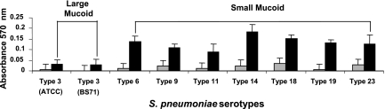
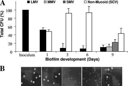
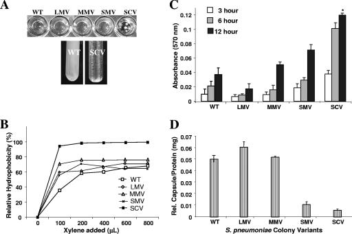
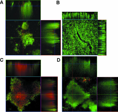
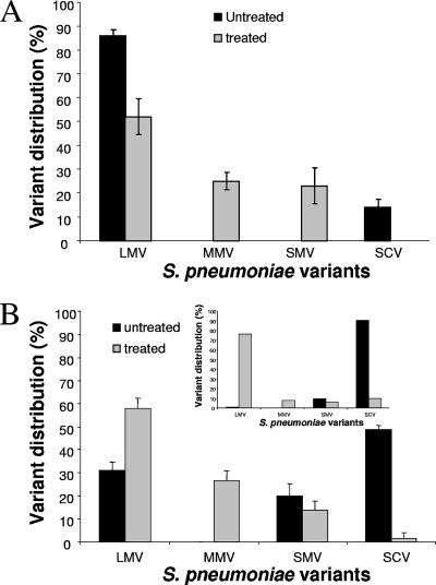
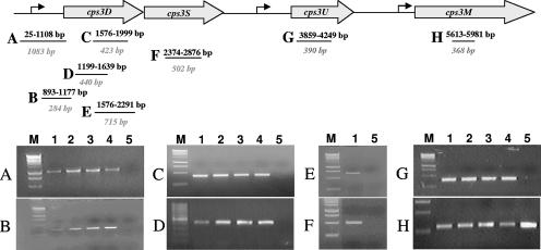
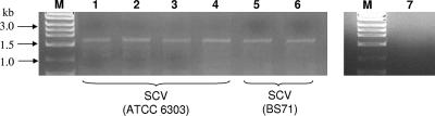
In this study, we report the isolation of colony morphology variants from Streptococcus pneumoniae serotype 3 biofilms. The colony variants differed in colony size (large, medium, and small) and their mucoid appearance on blood agar. The small nonmucoid variant (SCV) emerged during the initial attachment stage of S. pneumoniae biofilm formation and dominated over the course of biofilm growth. Mucoid variants appeared at later biofilm developmental stages. The reduction in colony size/mucoidy correlated with a decrease in capsule production and an increase in initial attachment. The large mucoid variant formed flat unstructured biofilms, failed to aggregate in liquid culture, and adhered poorly to solid surfaces. In contrast, SCVs autoaggregated in liquid culture, hyperadhered to solid surfaces, and formed biofilms with significant three-dimensional structure, mainly in the form of microcolonies. The variants showed similar antibiotic resistance/susceptibility based on a modified Kirby-Bauer test and when grown as biofilms. However, antimicrobial treatment of S. pneumoniae biofilms altered the colony variant's distribution and mainly affected the most interior areas of biofilm microcolonies. To further explore the nature of the variants, the capsule biosynthetic operon (cps3DSUM) was explored in greater detail. The genetic analysis indicated that the emergence of nonmucoid variants was due to a deletion comprising cps3DSU as well as additional genes upstream of the cps3 operon. Overall, our findings suggest that in vitro biofilm formation of S. pneumoniae serotype 3 coincides with the emergence of colony variants with distinct genotypic and phenotypic characteristics.


Figures

FIG. 1.
FIG. 1.
Initial biofilm attachment of different S. pneumoniae capsule serotypes. Various S. pneumoniae capsule serotypes were allowed to form biofilms in polystyrene microtiter plates for 6 (gray bars) and 12 h (black bars), after which time the attached biomass was stained with crystal violet, solubilized with ethanol, and subsequently measured at 570 nm. The capsule serotypes are grouped as large mucoid or small mucoid according to their appearance on blood agar.
FIG. 2.
FIG. 2.
Emergence of colony morphology variants over the course of S. pneumoniae serotype 3 biofilm development. (A) Distribution of colony variants was determined from total CFU, colony size, and mucoidy on blood agar. (B) Appearance of colony variants on solid medium (from left, inoculum, planktonic growth conditions after 1, 3, 6, and 9 days of biofilm growth).
FIG. 3.
FIG. 3.
Liquid culture phenotype, relative hydrophobicity (MATH), microtiter plate adhesion assay, and relative capsule concentration of S. pneumoniae WT and colony variants. (A) Hyperadherence and hyperautoaggregation of SCV in liquid cultures. The WT showed homogeneous growth in shaken liquid culture, as opposed to autoaggreation, seen for SCV. (B) Adherence to hexadecane as a measure of relative hydrophobicity (MATH) of colony morphology variants. Each datum point is based on the results from three replicate measurements. SCVs were significantly more hydrophobic than WT and the mucoid variants (P < 0.05). (C) Microtiter plate adhesion assay. SCV showed significantly greater biofilm adhesion than did WT, LMV, or MMV (P < 0.01). Initial attachment of S. pneumoniae colony variants was analyzed after 3, 6, and 12 h. (D) Relative capsule concentration of S. pneumoniae WT and colony variants as determined by using the Stains-All reagent.
FIG. 4.
FIG. 4.
Biofilm architecture of colony variants LMV and SCV and S. pneumoniae WT biofilms before and after treatment with tetracycline. (A) Three-day-old biofilm of SCV; (B) 3-day-old biofilm of LMV; (C) 6-day-old S. pneumoniae WT biofilm after treatment with tetracycline (10 μg/ml) for 12 h; (D) untreated 6-day-old S. pneumoniae WT biofilm. The CLSM images show the x-y and x-z planes. Flow cell experiments were performed in triplicate as described in Materials and Methods.
FIG. 5.
FIG. 5.
Colony variant distribution in S. pneumoniae biofilms is affected by treatment with tetracycline. Biofilms were grown in tube reactors for 1 day before treatment with tetracycline (10 μg/ml) for 6 h (A) or for 6 days before treatment with tetracycline (10 μg/ml) for 12 h (B). Inset: distribution of colony variants in 6-day-old S. pneumoniae WT biofilm grown in flow cells before and after treatment with tetracycline. Distribution of colony variants was assessed by total CFU, colony size, and mucoidy on blood agar. Experiments were performed in triplicate.
FIG. 6.
FIG. 6.
Mapping of the cps3DSU deletion in the genome of SCV by PCR. Genomic DNA of WT and mucoid variants was used as a control. The PCR product was amplified using the following primers: (A) cps3-promoter (1,083-bp PCR product); (B) cps3-promoter2 (284 bp); (C) cpsD (342 bp); (D) cps3Dint (440 bp); (E) cps3DS (715 bp); (F) cps3S (502 bp); (G) cps3U (390 bp); (H) cps3M (368 bp). Genomic DNAs isolated from S. pneumoniae wild type (6303), LMV, MMV, and SMV were used as controls. Lanes: 1, S. pneumoniae wild type (6303); 2, LMV; 3, MMV; 4, SMV; 5, SCV; M, DNA ladder (for PCR products shown in panels A to C and E to H, a 1-kb DNA ladder was used; for PCR products shown in panel D, a 100-bp DNA ladder was used).
FIG. 7.
FIG. 7.
Survey of SCVs obtained by PCR from independent biofilm growth experiments. Genomic DNA from SCVs obtained from four independent S. pneumoniae ATCC 6303 biofilm experiments (lanes 1 to 4) and from two independent S. pneumoniae BS71 biofilm experiments (lanes 5 and 6) was amplified using dexB and cps3M-del primers. Genomic DNA of S. pneumoniae ATCC 6303 grown planktonically was used as a control (lane 7). Lane M, 1-kb DNA ladder.

References

    1. Adamou, J. E., T. M. Wizemann, P. Barren, and S. Langermann. 1998. Adherence of Streptococcus pneumoniae to human bronchial epithelial cells (BEAS-2B). Infect. Immun. 66:820-822. - PMC - PubMed
    1. Allegrucci, M., F. Z. Hu, K. Shen, J. Hayes, G. D. Ehrlich, J. C. Post, and K. Sauer. 2005. Phenotypic characterization of Streptococcus pneumoniae biofilm development. J. Bacteriol. 188:2325-2335. - PMC - PubMed
    1. Austrian, R. 1981. Some observations on the pneumococcus and the current status of pneumococcal disease and its prevention. Rev. Infect. Dis. 3:S1-S17. - PubMed
    1. Bauer, A. W., D. M. Perry, and W. M. M. Kirby. 1959. Single disc antibiotic sensitivity testing of staphylococci. Arch. Int. Med. 104:208-216. - PubMed
    1. Boles, B. R., M. Thoendel, and P. K. Singh. 2004. Self-generated diversity produces “insurance effects” in biofilm communities. Proc. Natl. Acad. Sci. USA 101:16630-16635. - PMC - PubMed

Publication types

MeSH terms

Substances

LinkOut - more resources