. 2007 Feb;16(2):336-42.
doi: 10.1110/ps.062660907. Epub 2006 Dec 22.

Crystal structure of the BTB domain from the LRF/ZBTB7 transcriptional regulator

Affiliations

Crystal structure of the BTB domain from the LRF/ZBTB7 transcriptional regulator

Peter J Stogios et al. Protein Sci. 2007 Feb.

Abstract

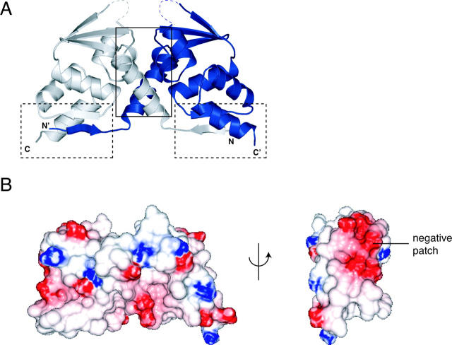
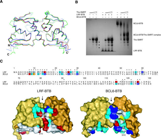
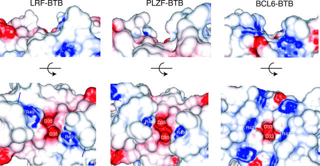
BTB-zinc finger (BTB-ZF) proteins are transcription regulators with roles in development, differentiation, and oncogenesis. In these proteins, the BTB domain (also known as the POZ domain) is a protein-protein interaction motif that contains a dimerization interface, a possible oligomerization surface, and surfaces for interactions with other factors, including nuclear co-repressors and histone deacetylases. The BTB-ZF protein LRF (also known as ZBTB7, FBI-1, OCZF, and Pokemon) is a master regulator of oncogenesis, and represses the transcription of a variety of important genes, including the ARF, c-fos, and c-myc oncogenes and extracellular matrix genes. We determined the crystal structure of the BTB domain from human LRF to 2.1 A and observed the canonical BTB homodimer fold. However, novel features are apparent on the surface of the homodimer, including differences in the lateral groove and charged pocket regions. The residues that line the lateral groove have little similarity with the equivalent residues from the BCL6 BTB domain, and we show that the 17-residue BCL6 Binding Domain (BBD) from the SMRT co-repressor does not bind to the LRF BTB domain.


Figures

Figure 1.
Figure 1.
Structure of the LRF BTB domain. (A) Ribbon representation. (Dashed lines) Regions of the turn between α3 and β4 with missing electron density; (solid box) open dimerization interface; (dashed boxes) closed dimerization interfaces. (B) Electrostatic surface representation of the LRF-BTB homodimer, shown in two views. A large negatively charged patch is labeled.
Figure 2.
Figure 2.
Comparison of the LRF and BCL6 BTB domains. (A) Superposition of Cα atoms of the LRF, PLZF, and BCL6 BTB homodimers (PDB ID codes 2NN2, 1BUO, 1R29). (Red) LRF, (blue) PLZF, (green) BCL6. (B) Binding of SMRT-BBD to BTB domains. LRF and BCL6 BTB domain were titrated with increasing amounts of Thioredoxin-SMRT (Thx-SMRT) fusion protein and separated by native gel electrophoresis. Lanes 13 contained Thx-SMRT alone (2.5, 8, and 13 μg), lanes 47 and 811 contained 10 μg LRF-BTB or BCL6-BTB, respectively, and 0, 2.5, 8, or 13 μg of Thx-SMRT. (Asterisks) Three minor impurities in the Thx-SMRT sample. (C) Comparison of the lateral grooves of the LRF and BCL6 BTB domains. A sequence alignment of LRF and BCL6-BTB is shown. (Asterisks) Residues located in the lateral groove region. Lateral groove residues that show non-conservative substitutions between the two BTB domains are shaded by residue type: (blue) basic, (red) acidic, (gray) hydrophobic, (cyan) polar, and (yellow) sulfur-containing. Conservative substitutions are not shaded, except for His 116/Ala 118 (boxed). Residues are numbered according to the LRF sequence. The region of the disordered loop in the LRF structure is underlined. The structures of the LRF and BCL6 BTB domains are shown in surface representation below the sequence alignment, with the lateral groove residues colored as in the alignment. Residues that have the largest buried surface areas in the BCL6-SMRT complex, and their equivalents in LRF, are labeled. Residues not involved in the lateral groove are colored shades of yellow. (Arrow) The charged pocket region of the BTB domains.
Figure 3.
Figure 3.
Comparison of the charged pocket of the LRF, PLZF, and BCL6 BTB homodimers (PDB ID codes 2NN2, 1BUO, 1R29), shown as electrostatic surface representations. Two views are shown, the first in the same perspective as Fig. 2, the second rotated 90° toward the reader. Residues Asp 33/35 and Arg 47/49 are labeled.

References

    1. Ahmad, K.F., Engel, C.K., and Privé, G.G. 1998. Crystal structure of the BTB domain from PLZF. Proc. Natl. Acad. Sci. 95: 12123–12128. - PMC - PubMed
    1. Ahmad, K.F., Melnick, A., Lax, S., Bouchard, D., Liu, J., Kiang, C.L., Mayer, S., Takahashi, S., Licht, J.D., and Privé, G.G. 2003. Mechanism of SMRT corepressor recruitment by the BCL6 BTB domain. Mol. Cell 12: 1551–1564. - PubMed
    1. Collins, T., Stone, J.R., and Williams, A.J. 2001. All in the family: The BTB/POZ, KRAB, and SCAN domains. Mol. Cell. Biol. 21: 3609–3615. - PMC - PubMed
    1. David, G., Alland, L., Hong, S.H., Wong, C.W., DePinho, R.A., and Dejean, A. 1998. Histone deacetylase associated with mSin3A mediates repression by the acute promyelocytic leukemia-associated PLZF protein. Oncogene 16: 2549–2556. - PubMed
    1. Davies, J.M., Hawe, N., Kabarowski, J., Huang, Q.H., Zhu, J., Brand, N.J., Leprince, D., Dhordain, P., Cook, M., and Morriss-Kay, G., et al. 1999. Novel BTB/POZ domain zinc-finger protein, LRF, is a potential target of the LAZ-3/BCL-6 oncogene. Oncogene 18: 365–375. - PubMed

Publication types

MeSH terms

Associated data

LinkOut - more resources