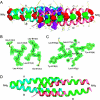
Self-assembly of coiled-coil tetramers in the 1.40 A structure of a leucine-zipper mutant
- PMID: 17189475
- PMCID: PMC2203300
- DOI: 10.1110/ps.062590807
Self-assembly of coiled-coil tetramers in the 1.40 A structure of a leucine-zipper mutant
Abstract
The hydrophobic core of the GCN4 leucine-zipper dimerization domain is formed by a parallel helical association between nonpolar side chains at the a and d positions of the heptad repeat. Here we report a self-assembling coiled-coil array formed by the GCN4-pAe peptide that differs from the wild-type GCN4 leucine zipper by alanine substitutions at three charged e positions. GCN4-pAe is incompletely folded in normal solution conditions yet self-assembles into an antiparallel tetraplex in crystals by formation of unanticipated hydrophobic seams linking the last two heptads of two parallel double-stranded coiled coils. The GCN4-pAe tetramers in the lattice associate laterally through the identical interactions to those in the intramolecular dimer-dimer interface. The van der Waals packing interaction in the solid state controls extended supramolecular assembly of the protein, providing an unusual atomic scale view of a mesostructure.
Figures


Similar articles
-
Antiparallel four-stranded coiled coil specified by a 3-3-1 hydrophobic heptad repeat.Structure. 2006 Feb;14(2):247-55. doi: 10.1016/j.str.2005.10.010. Structure. 2006. PMID: 16472744 Free PMC article.
-
A parallel coiled-coil tetramer with offset helices.Biochemistry. 2006 Dec 26;45(51):15224-31. doi: 10.1021/bi061914m. Epub 2006 Nov 29. Biochemistry. 2006. PMID: 17176044
-
A switch between two-, three-, and four-stranded coiled coils in GCN4 leucine zipper mutants.Science. 1993 Nov 26;262(5138):1401-7. doi: 10.1126/science.8248779. Science. 1993. PMID: 8248779
-
Pharmacological interference with protein-protein interactions mediated by coiled-coil motifs.Handb Exp Pharmacol. 2008;(186):461-82. doi: 10.1007/978-3-540-72843-6_19. Handb Exp Pharmacol. 2008. PMID: 18491064 Review.
-
Boehringer Mannheim award lecture 1995. La conference Boehringer Mannheim 1995. De novo design of alpha-helical proteins: basic research to medical applications.Biochem Cell Biol. 1996;74(2):133-54. doi: 10.1139/o96-015. Biochem Cell Biol. 1996. PMID: 9213423 Review.
Cited by
-
The effects of pK(a) tuning on the thermodynamics and kinetics of folding: design of a solvent-shielded carboxylate pair at the a-position of a coiled-coil.Biophys J. 2010 Oct 6;99(7):2299-308. doi: 10.1016/j.bpj.2010.07.059. Biophys J. 2010. PMID: 20923665 Free PMC article.
-
Cohesin interaction with centromeric minichromosomes shows a multi-complex rod-shaped structure.PLoS One. 2008 Jun 11;3(6):e2453. doi: 10.1371/journal.pone.0002453. PLoS One. 2008. PMID: 18545699 Free PMC article.
-
Two Engineered OBPs with opposite temperature-dependent affinities towards 1-aminoanthracene.Sci Rep. 2018 Oct 4;8(1):14844. doi: 10.1038/s41598-018-33085-8. Sci Rep. 2018. PMID: 30287882 Free PMC article.
-
Confirmation of Bioinformatics Predictions of the Structural Domains in Honeybee Silk.Polymers (Basel). 2018 Jul 16;10(7):776. doi: 10.3390/polym10070776. Polymers (Basel). 2018. PMID: 30960701 Free PMC article.
-
Essential role of coiled coils for aggregation and activity of Q/N-rich prions and PolyQ proteins.Cell. 2010 Dec 23;143(7):1121-35. doi: 10.1016/j.cell.2010.11.042. Cell. 2010. PMID: 21183075 Free PMC article.
References
-
- Betz, S.F., Bryson, J.W., and DeGrado, W.F. 1995. Native-like and structurally characterized designed α-helical bundles. Curr. Opin. Struct. Biol. 5: 457–463. - PubMed
-
- Brunger, A.T., Adams, P.D., Clore, G.M., DeLano, W.L., Gros, P., Grosse-Kunstleve, R.W., Jiang, J.S., Kuszewski, J., Nilges, M., and Pannu, N.S., et al. 1998. Crystallography & NMR system: A new software suite for macromolecular structure determination. Acta Crystallogr. D Biol. Crystallogr. 54: 905–921. - PubMed
-
- Bryson, J.W., Betz, S.F., Lu, H.S., Suich, D.J., Zhou, H.X., O'Neil, K.T., and DeGrado, W.F. 1995. Protein design: A hierarchic approach. Science 270: 935–941. - PubMed
-
- Crick, F.H.C. 1953. The packing of α-helices: Simple coiled-coils. Acta Crystallogr. 6: 689–697.
Publication types
MeSH terms
Substances
Grants and funding
LinkOut - more resources
Full Text Sources
Molecular Biology Databases

