

Tension band stabilisation of acetabular physeal fractures in four kittens
- PMID: 17189710
- PMCID: PMC10822614
- DOI: 10.1016/j.jfms.2006.10.004
Tension band stabilisation of acetabular physeal fractures in four kittens
Abstract
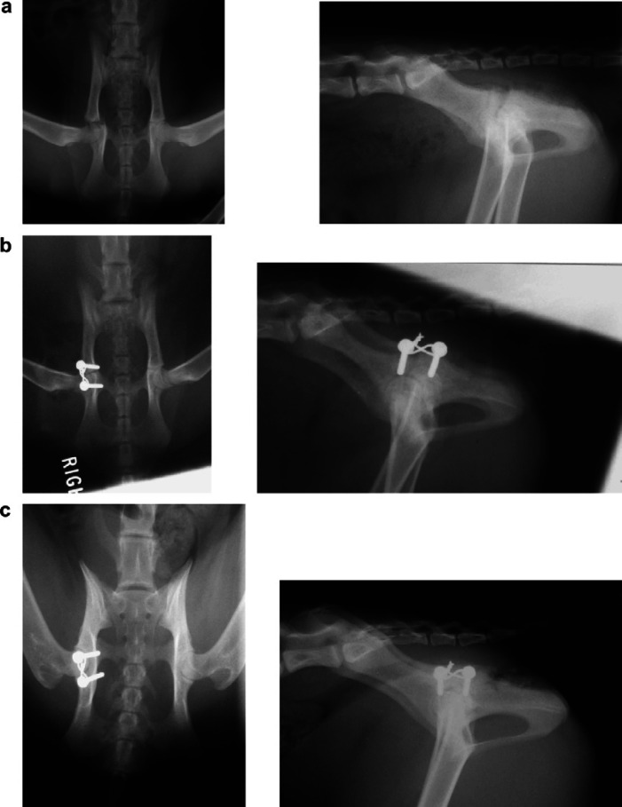
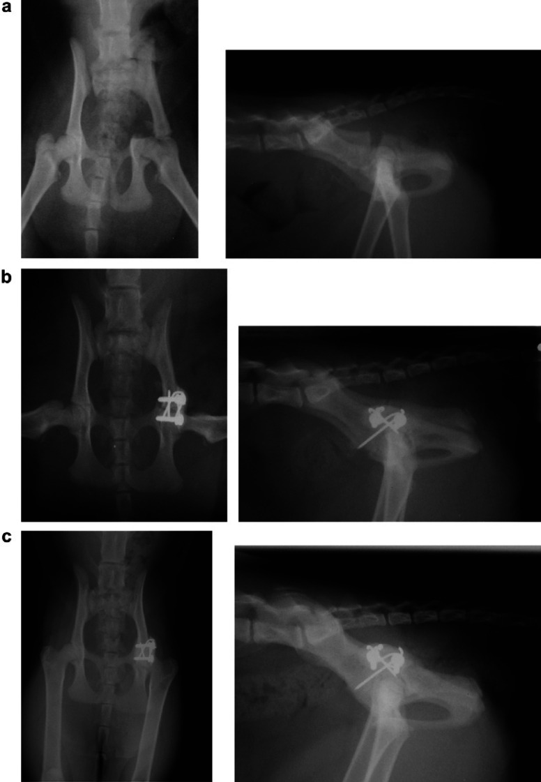
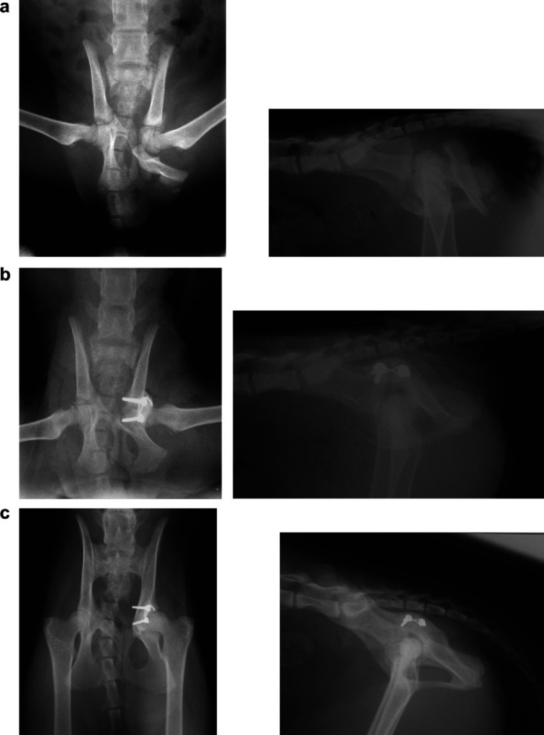
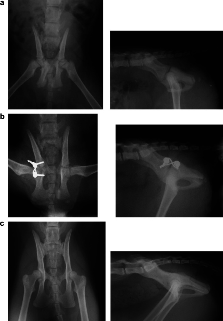
The surgical repair of acetabular physeal fractures in four kittens using a screw and tension band technique is reported. This was an appropriate method for restoring articular congruency and improving pelvic alignment. All cases had an excellent outcome and full limb use following fracture repair. In kittens younger than 12 weeks, there is a possibility of premature fusion of the acetabular bone resulting in development of a deformed, shallow acetabulum and hip subluxation. However, surgery is still justified when there is pelvic canal narrowing to decrease the risk of future defecatory problems. Early implant removal in such young kittens may decrease the severity of deformity caused by premature physeal closure. In kittens of 16 weeks or older, the prognosis is good for normal acetabular development and implant removal is not necessary.
Figures
Similar articles
-
Management and long-term outcome of pelvic fractures: a retrospective study of 43 cats.J Feline Med Surg. 2017 Jan;19(1):36-41. doi: 10.1177/1098612X15606958. Epub 2016 Jul 10. J Feline Med Surg. 2017. PMID: 26445978 Free PMC article.
-
Traumatic physeal fractures in cats: a review of 36 cases (2010-2020).J Feline Med Surg. 2022 Feb;24(2):98-106. doi: 10.1177/1098612X211005886. Epub 2021 Apr 13. J Feline Med Surg. 2022. PMID: 33847538 Free PMC article.
-
Tension band wire for acetabular fracture fixation: a technical note.J Orthop Surg (Hong Kong). 2011 Dec;19(3):386-8. doi: 10.1177/230949901101900328. J Orthop Surg (Hong Kong). 2011. PMID: 22184179
-
Minimally invasive repair of sacroiliac luxation in small animals.Vet Clin North Am Small Anim Pract. 2012 Sep;42(5):1069-77, viii. doi: 10.1016/j.cvsm.2012.06.005. Vet Clin North Am Small Anim Pract. 2012. PMID: 23040309 Review.
-
Pelvic fractures in children (pelvic ring and acetabulum).Orthop Traumatol Surg Res. 2020 Feb;106(1S):S125-S133. doi: 10.1016/j.otsr.2019.05.017. Epub 2019 Sep 11. Orthop Traumatol Surg Res. 2020. PMID: 31521559 Review.
Cited by
-
A Bilateral Acetabular Physeal Fracture Treated with External Fixation in an Immature Cat.Animals (Basel). 2024 Jan 25;14(3):379. doi: 10.3390/ani14030379. Animals (Basel). 2024. PMID: 38338023 Free PMC article.
-
Management of pelvic trauma: neurological damage, urinary tract disruption and pelvic fractures.J Feline Med Surg. 2011 May;13(5):347-61. doi: 10.1016/j.jfms.2011.03.011. J Feline Med Surg. 2011. PMID: 21515222 Free PMC article. Review.
-
Management and long-term outcome of pelvic fractures: a retrospective study of 43 cats.J Feline Med Surg. 2017 Jan;19(1):36-41. doi: 10.1177/1098612X15606958. Epub 2016 Jul 10. J Feline Med Surg. 2017. PMID: 26445978 Free PMC article.
-
Outcome of surgical stabilisation of acetabular fractures in 16 cats.J Feline Med Surg. 2019 Jun;21(6):520-528. doi: 10.1177/1098612X18788165. Epub 2018 Aug 3. J Feline Med Surg. 2019. PMID: 30074433 Free PMC article.
-
Composite fixation of comminuted ilial wing fractures in cats: three cases.J Feline Med Surg. 2011 May;13(5):376-82. doi: 10.1016/j.jfms.2011.03.013. J Feline Med Surg. 2011. PMID: 21515224 Free PMC article.
References
-
- Beaver D.P., Lewis D.D., Lanz O.I., Madison J.B., Kubilis P.S. Evaluation of four interfragmentary Kirschner wire configurations as a component of screw/wire/polymethylmethacrylate fixation for acetabular fractures in dogs, Journal of the American Animal Hospital Association 36, 2000, 456–462. - PubMed
-
- Bookbinder P.F., Flanders J.A. Characteristics of pelvic fracture in the cat, Veterinary Comparative Orthopaedics and Traumatology 5, 1992, 122–127.
-
- Brinker W.O., Piermattei D.L., Flo G.L. Fractures of the pelvis. Brinker W.O., Piermattei D.L., Flo G.L. Brinker, Piermattei and Flo's Handbook of Small Animal Orthopaedics and Fracture Repair, 3rd edn, 1997a, W.B. Saunders Co: Philadelphia, 395–421.
-
- Brinker W.O., Piermattei D.L., Flo G.L. Fractures in growing animals. Brinker W.O., Piermattei D.L., Flo G.L. Brinker, Piermattei and Flo's Handbook of Small Animal Orthopaedics and Fracture Repair, 3rd edn, 1997b, W.B. Saunders Co: Philadelphia, 676–685.
-
- Brown R.E. A surgical approach to the coxo-femoral joint of dogs, North American Veterinarian 34, 1953, 420–422.
MeSH terms
LinkOut - more resources
Full Text Sources
Medical
