. 2007 Jan;11(1):9-23.
doi: 10.1016/j.ccr.2006.10.019. Epub 2006 Dec 28.

Mad2 overexpression promotes aneuploidy and tumorigenesis in mice

Affiliations

Mad2 overexpression promotes aneuploidy and tumorigenesis in mice

Rocío Sotillo et al. Cancer Cell. 2007 Jan.

Abstract

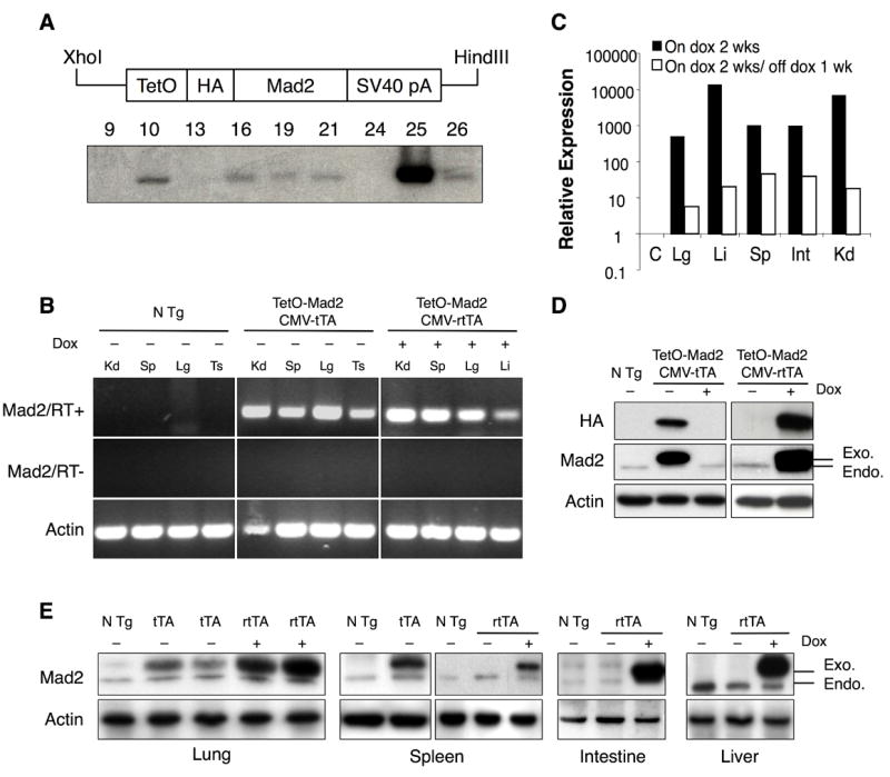
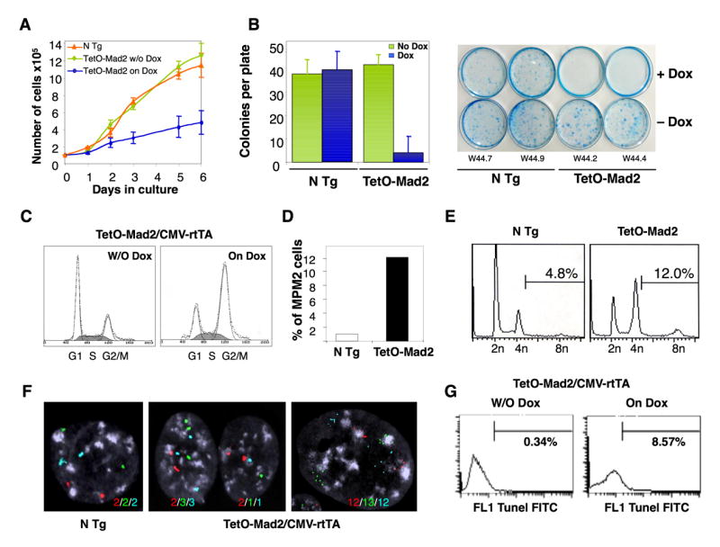
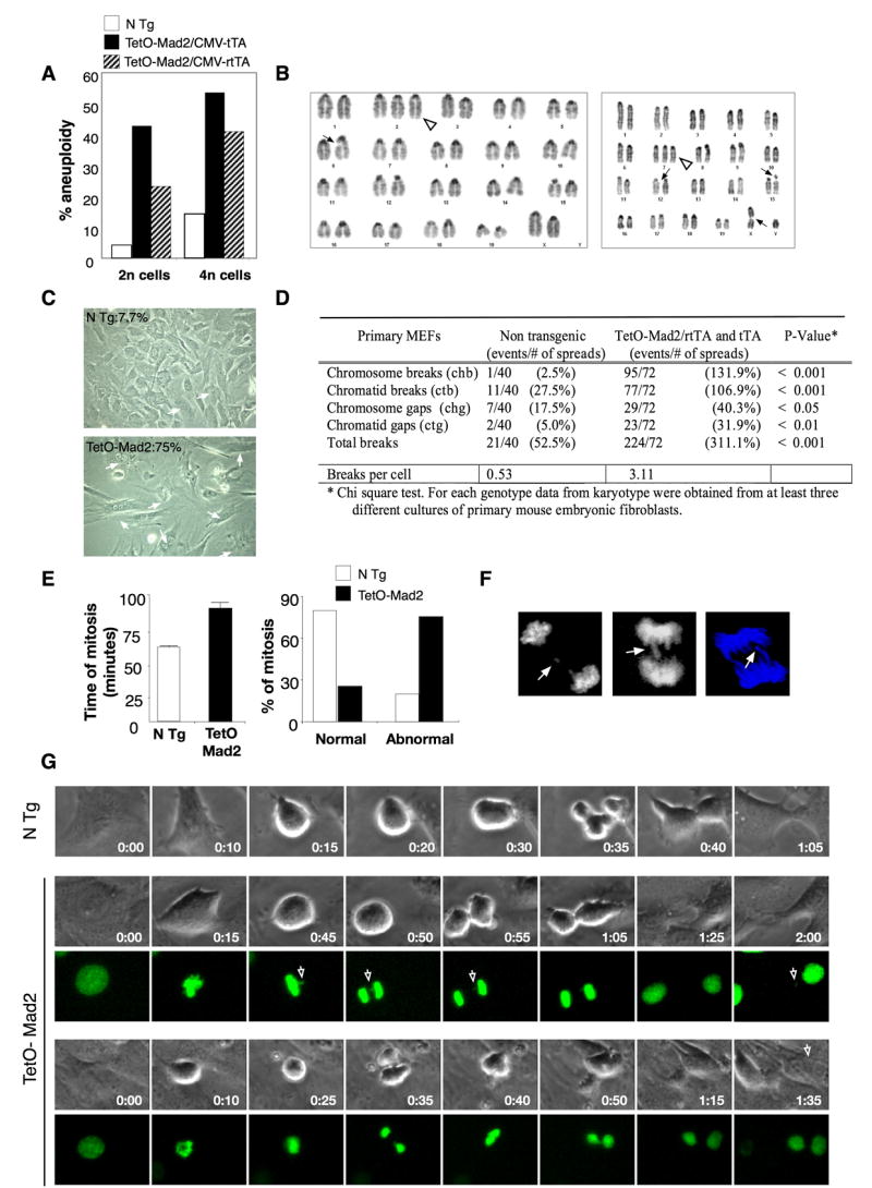
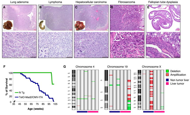
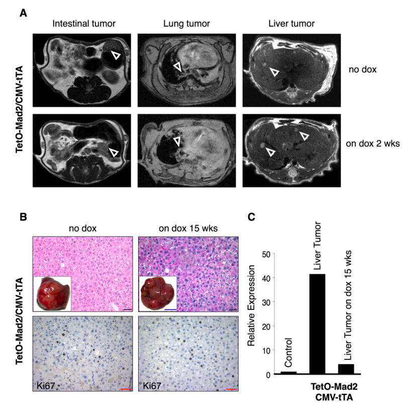
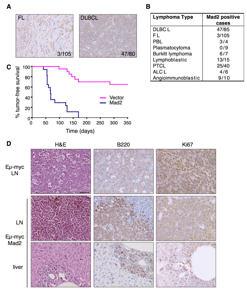
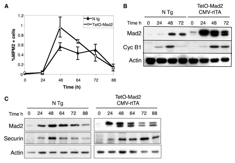
Mad2 is an essential component of the spindle checkpoint that blocks activation of Separase and dissolution of sister chromatids until microtubule attachment to kinetochores is complete. We show here that overexpression of Mad2 in transgenic mice leads to a wide variety of neoplasias, appearance of broken chromosomes, anaphase bridges, and whole-chromosome gains and losses, as well as acceleration of myc-induced lymphomagenesis. Moreover, continued overexpression of Mad2 is not required for tumor maintenance, unlike the majority of oncogenes studied to date. These results demonstrate that transient Mad2 overexpression and chromosome instability can be an important stimulus in the initiation and progression of different cancer subtypes.


Figures

Figure 1
Figure 1
Mad2 transgene and expression pattern. (A) Construct used to generate the tetracycline operator-regulated Mad2 (TetO-Mad2) responder mice. TetO, tetracycline operator; HA, hemaglutinin; SV40 pA, SV40 gene polyadenylation sequence (upper panel). Southern blot of genomic DNA from different founders (lower panel). (B) RT-PCR from different tissues of non-transgenic, TetO-Mad2/CMV-tTA and TetO-Mad2/CMV-rtTA mice, the last ones exposed to doxycycline in the feed from 4 weeks to harvest at 8 weeks. PCR reactions were carried out in the presence (top) and absence (middle) of RT and products were visualized after electrophoresis in a 2% agarose gel. Amplification of actin mRNA by RT-PCR confirmed the presence of RNA in all samples. (C) Quantitative RT-PCR analysis of transgene expression in bitransgenic (Tet-On) mice on doxycycline and after doxycycline withdrawal (C= control, Kd=Kidney, Sp=spleen, Li=Liver, Lg=lung, Ts=Testis Int=intestine). (D) Western blot analysis for HA tag and Mad2 of non transgenic (N Tg) and TetO-Mad2/CMV-tTA MEFs (left panel) and TetO-Mad2/CMV-rtTA MEFs (right panel). Cells were maintained with (+) or without (−) doxycycline for 24h. (E) Western blot analysis showing Mad2 levels in different tissues from TetO-Mad2/CMV-tTA mice (lung and spleen) and TetO-Mad2/CMV-rtTA mice with and without doxycycline treatment (lung, spleen, intestine and liver). N Tg: non transgenic mice, tTA: is TetO-Mad2/CMV-tTA mice and rtTA: TetO-Mad2/CMV-rtTA mice. Anti-actin blots are shown as a loading control.
Figure 2
Figure 2
Growth properties of TetO-Mad2/CMV-rtTA MEFs. (A) Proliferation of early passage MEFs with or without the addition of doxycycline. (B) Plating efficiency of Mad2 wild type and TetO-Mad2/CMV-rtTA MEFs with and without doxycycline treatment. (C) Cell cycle profile of asynchronous cultures at passage 2. (D) Percentage of cells positive for MPM2 as a marker of mitosis. (E) FACS analysis of DNA content profile of asynchronously growing non transgenic and TetO-Mad2/CMV-rtTA MEFs on doxycycline media. (F) FISH analysis on MEFs using centromeric probes for chromosomes 12 (red), 16 (green) and 17 (blue) showing a non transgenic cell (left panel), a binucleated cell (middle panel) and a mononucleated cell with more than 4N (right panel). (G) TUNEL assay of TetO-Mad2/CMV-rtTA MEFs with and without doxycycline for 48 hours.
Figure 3
Figure 3
Overexpression of Mad2 leads to chromosomal instability. (A) Percentage of aneuploidy in the 2n and 4n population in TetO-Mad2 overexpressing MEFs in the Tet-On and Tet-Off systems. (B) Karyotype of a Tet-On cell with an extra chromosome (2) (white triangle) and a chromatid break (6) (ctb) (left panel), and karyotype of another cell with an extra chromosome 7 (white triangle), a ctb (X), ctb (15) and chromatid gap (ctg) (12) (right panel). (C) Representative picture of Tet-On P2 MEFs in culture showing binucleated cells (white arrows). (D) Number of chromosomal breaks on primary MEFs. (E, right panel) Time of mitosis of non transgenic (n = 98) and TetO-Mad2 (n = 90) MEFs was followed by time-lapse microscopy. Mean time of total mitosis is shown. (E, left panel) Percentage of cells with normal or abnormal mitosis (binucleated cells, furrow regression, chromosome bridges and mitotic catastrophe) as assessed by time-lapse microscopy. (F) Evidence of lagging chromosomes (top panel) and chromosome bridges (bottom 2 panels). (G) Time-lapse micrography of non transgenic and TetO-Mad2 MEFs. (Upper) N Tg cell entering mitosis at T=0 min and completing cytokinesis by 1 hour. (Middle) Representative cell overexpressing Mad2 with a chromosome bridge (white arrow) stays longer in mitosis and exits at 1hr 25 min with a missegregated chromosome (arrow). (Lower) Example of a cell with a chromosome bridge that suffers furrow regression and exits mitosis as a binucleated cell.
Figure 4
Figure 4
Tumor susceptibility in TetO-Mad2/CMV-tTA mice. Micrography of H&E stainings of a lung adenoma (A); a metastatic lymphoma in the colon (B); an hepatocellular carcinoma (C); a fibrosarcoma in the skin (D) and a dysplasia of the fallopian tubes (E). Insets on A,B,C: macroscopic pictures of the corresponding tissues. Inset B, C lower panel: detail of abnormal mitoses; Blue bar 1cm. Red bar 80μm and black bar 100 μm. (F) Survival curve of TetO-Mad2/CMV-tTA mice where blue line represents TetO-Mad2/CMV-tTA mice (n=40) and green line are non transgenic littermates (n=15). (G) Chromosomal abnormalities detected by comparative genomic hybridization array analysis of TetO-Mad2/CMV-tTA normal livers and liver tumors compared to a wild type liver showing amplification and deletion of specific regions as well as a whole chromosome gain (X).
Figure 5
Figure 5
Overexpression of Mad2 is not required for tumor maintenance. (A) Axial MR images of the abdomen and the lungs of bitransgenic mice on normal diet (Mad2 on) and after 2 weeks on doxycycline food (Mad2 off) showing the presence of tumors (white arrows). (B) H&E (top) and Ki67 staining (bottom) of a hepatoma in a bitransgenic TetO-Mad2/CMV-tTA mouse untreated (Mad2 on) (left) and a mouse fed doxycycline for 15 weeks after confirming the presence of a tumor by MRI. (C) QRT-PCR confirming the downregulation of Mad2 in the tumor. Black bar 100μm, red bar 20μm and blue bar 1cm.
Figure 6
Figure 6
Mad2 overexpression, a common feature of certain human lymphomas, accelerates myc-driven lymphomagenesis. (A) Immunohistochemical evaluation of Mad2 expression in human FL and DLBCL. Two representative positive cases are shown. (B) Results of Mad2 immunohistochemistry analysis in multiple human lymphoma subtypes. (C) Tumor free survival curve of animals transplanted with Eμ-myc/vector vs. Eμ-myc/Mad2 HSCs showing statistically significant acceleration of tumor initiation mediated by Mad2 induction. (D) Histological and immunohistochemical evaluation of Eμ-myc and Eμ-myc/Mad2-derived lymphomas (LN) and liver metastases.
Figure 7
Figure 7
Mitotic progression of in vitro stimulated lymphocytes. (A) Percentage of lymphocytes positive for MPM2 as a marker of mitosis. (B) Western blot analysis of in vitro stimulated lymphocytes isolated from spleen of non transgenic mice and TetO-Mad2/CMV-rtTA mice in the presence of doxycycline showing stabilization of cyclin B1 in the Mad2 overexpressing cells as compared to the non transgenic cells (C) Western blot analysis of in vitro stimulated lymphocytes showing stabilization of securin in the TetO-Mad2/CMV-rtTA cells compared to the non transgenic control.

Comment in

References

    1. Adams JM, Harris AW, Pinkert CA, Corcoran LM, Alexander WS, Cory S, Palmiter RD, Brinster RL. The c-myc oncogene driven by immunoglobulin enhancers induces lymphoid malignancy in transgenic mice. Nature. 1985;318:533–538. - PubMed
    1. Alizadeh AA, Eisen MB, Davis RE, Ma C, Lossos IS, Rosenwald A, Boldrick JC, Sabet H, Tran T, Yu X, et al. Distinct types of diffuse large B-cell lymphoma identified by gene expression profiling. Nature. 2000;403:503–511. - PubMed
    1. Babu JR, Jeganathan KB, Baker DJ, Wu X, Kang-Decker N, van Deursen JM. Rae1 is an essential mitotic checkpoint regulator that cooperates with Bub3 to prevent chromosome missegregation. J Cell Biol. 2003;160:341–353. - PMC - PubMed
    1. Baker DJ, Jeganathan KB, Cameron JD, Thompson M, Juneja S, Kopecka A, Kumar R, Jenkins RB, de Groen PC, Roche P, van Deursen JM. BubR1 insufficiency causes early onset of aging-associated phenotypes and infertility in mice. Nat Genet. 2004;36:744–749. - PubMed
    1. Bharadwaj R, Yu H. The spindle checkpoint, aneuploidy, and cancer. Oncogene. 2004;23:2016–2027. - PubMed

Publication types

LinkOut - more resources