. 2007 Feb;41(2):206-21.
doi: 10.1016/j.ymeth.2006.07.033.

dSLAM analysis of genome-wide genetic interactions in Saccharomyces cerevisiae

Affiliations

dSLAM analysis of genome-wide genetic interactions in Saccharomyces cerevisiae

Xuewen Pan et al. Methods. 2007 Feb.

Abstract

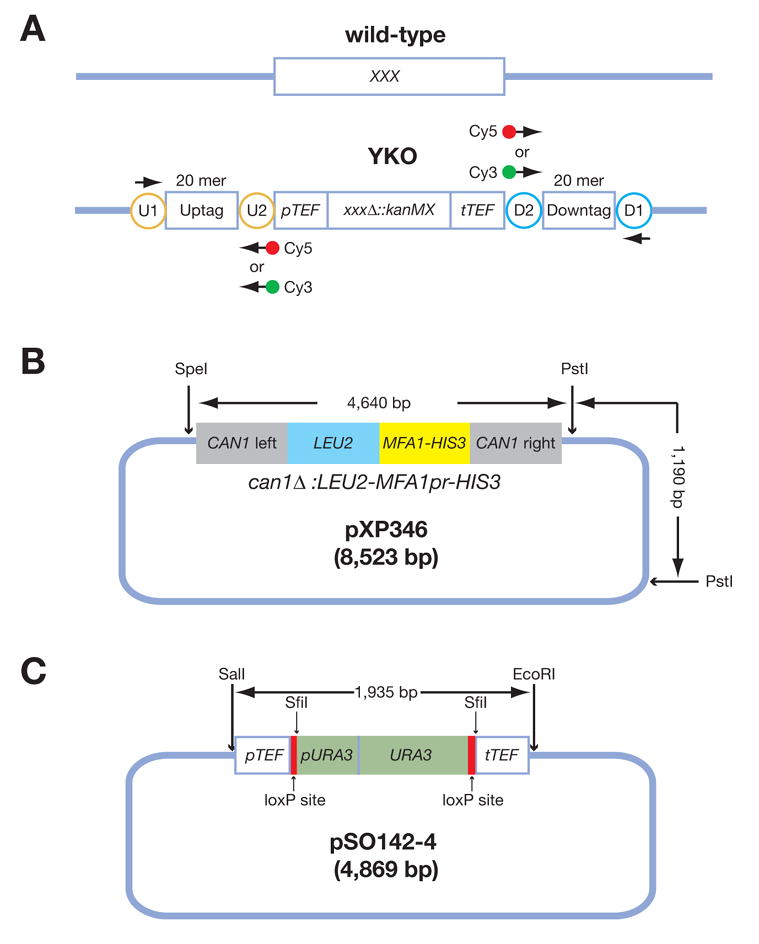
Analysis of genetic interactions has been extensively exploited to study gene functions and to dissect pathway structures. One such genetic interaction is synthetic lethality, in which the combination of two non-lethal mutations leads to loss of organism viability. We have developed a dSLAM (heterozygote diploid-based synthetic lethality analysis with microarrays) technology that effectively studies synthetic lethality interactions on a genome-wide scale in the budding yeast Saccharomyces cerevisiae. Typically, a query mutation is introduced en masse into a population of approximately 6000 haploid-convertible heterozygote diploid Yeast Knockout (YKO) mutants via integrative transformation. Haploid pools of single and double mutants are freshly generated from the resultant heterozygote diploid double mutant pool after meiosis and haploid selection and studied for potential growth defects of each double mutant combination by microarray analysis of the "molecular barcodes" representing each YKO. This technology has been effectively adapted to study other types of genome-wide genetic interactions including gene-compound synthetic lethality, secondary mutation suppression, dosage-dependent synthetic lethality and suppression.


Figures

Figure 1
Figure 1. Simplified structural diagrams for the YKO construct, pXP346, and pSO142-4
(A) A diagram for the yeast knockout construct. Each YKO consists of a kanMX module that confers resistance to the antibiotic G418 flanked by unique 20 mer “molecular barcodes” or “Tags” called the “Uptag” and “Downtag.” All “Uptags” are themselves flanked by a common set of priming sites (U1 and U2 within the orange circles) and all “Downtags” are flanked by another set of common priming sites (D1 and D2 within the cyan circles). These common priming sites allow for PCR amplification and microarray analysis of all Uptags or all Downtags in a population. “XXX” stands for any yeast gene. (B) A simplified diagram of pXP346, which contains the can1Δ::LEU2-MFA1pr-HIS3 reporter. This plasmid needs to be digested with SpeI and PstI to release the reporter for integration into the CAN1 locus of the yeast genome via homologous recombination. (C) A diagram of pSO142-4, which contains the URA3-loxP cassette. URA3 targeting construct pSO142 can be integrated into the existing kanMX4-marked YKO strains based on its sequence homology to the promoter and terminator sequences, derived from the Ashbya gossypii TEF gene. URA3, driven by its own promoter, is flanked by loxP sites allowing subsequent excision/marker swaps upon expression of Cre recombinase, increasing the flexibility of the marker for future manipulations. The SwaI and SfiI restriction sites were introduced for diagnostic purposes. In the first step, a loxP-URA3 cassette containing SwaI and SfiI sites was constructed using PCR. The loxP-URA3 cassette was amplified as two separate fragments, loxP-partial URA3 (loxP-URA3’) and partial URA3-loxP (‘URA3-loxP). The loxP, SwaI and SfiI sequences (introduced on primers) were fused to the 5′ and 3′ ends of the URA3 fragment via three sequential rounds of PCR. The URA3 fragment was amplified from pRS406. The loxP sequence was obtained from (25). Both PCR products were then co-transformed into a nej1 ::kanMX4 strain. The goal was to have the two PCR products homologously replace the kan ORF sequence. Integrative transformation was made possible by the following features on the two PCR products: loxP-URA3 contains 45 bp sequence homology to the TEF promoter, while URA3-loxP contains 48 bp sequence homology to the TEF terminator. The 3′ end of loxP-URA3’ and the 5′ end of ‘URA3-loxP overlap by 111 bp. nej1_ was used to decrease the efficiency of non-homologous end-joining. We first selected for Ura+ transformants. Next, we assayed and identified Ura+ transformants that are G418s, indicating that URA3 gene was integrated into kanMX4; one such strain was YSO205. In the second step, genomic DNA isolated from YSO205 was used as template to amplify the pTEF-loxP-URA3-loxP-tTEF fragment (1965 bp) using primers U2 and D2. The PCR products were then digested with SalI and EcoRI and cloned into pBSIIKS(−). Primer U2 contains a SalI site, while D2 contains EcoRI site. Note: The plasmid backbones in both panels B and C are not drawn to scale.
Figure 2
Figure 2. A flowchart for a dSLAM screen
Step 1. A query construct yfgΔ::URA3 is PCR-amplified and transformed en masse into a pool of haploid convertible heterozygote diploid YKOs. Step 2. The resultant double mutant pool is sporulated. Step 3. Haploid experiment (double mutants) and control (single and double mutants) pools are freshly and independently generated from the same sporulation culture by plating on the haploid selection media MM-Ura and MM, respectively. Step 4. Genomic DNA samples are prepared from these two haploid pools. Step 5. The TAGs from both pools are PCR-amplified with differentially labeled primers (Cy3 and Cy5). Step 6. The PCR-amplified dye-labeled TAGs are hybridized to a TAG-array and analyzed for the relative abundance of each TAG in both control and experiment pools. “YFG” stands for “Your Favorite Gene;” “XXX” stands for any gene in the yeast genome; “MM” stands for the haploid selection “Magic Medium” (see Table 3).
Figure 3
Figure 3. A flowchart for high throughput yeast transformation and random spore analysis of synthetic lethality interactions
Step 1. Individual haploid-convertible heterozygote diploid YKOs are picked, inoculated onto OmniTrays containing solid YPD plus G418 (200 μg/ml) in a 96-well format, and incubated at 30°C for overnight. Step 2. These strains are inoculated into fresh liquid YPD (100 μl/well) in a shallow 96-well plate with a 96-pin replicator and incubated at 30°C overnight. Step 3. 25 μl of each of the overnight cultures is transferred to a deep 96-well plate containing fresh liquid YPD (1 ml/well). The plate is shaken at 200 rpm at 30°C for 4 hours. Step 3. Cells are pelleted, washed once in 0.1 M LiOAc (200 μl/well), and transferred to a shallow 96-well plate as a cell suspension in 0.1 M LiOAc (100 μl/well). Step 4. After incubation with the transformation mixture at 30°C for 30 minutes and subsequently at 42°C for 13 minutes, cells are pelleted, resuspended in 5 mM CaCl2 (30 μl/well) for 5–15 minutes, transferred row by row onto solid SC-Ura OmniTray plates, and incubated at 30°C for 2 days. Step 5. Two isolated colonies are picked for each strain and patched onto fresh SC-Ura plates back into the original 96-well format and incubated at 30°C overnight. Step 6. Cells are transferred to solid sporulation medium with a 96-pin replicator and incubated at room temperature (22 to 25°C) for 5 days. Step 7. The sporulated cultures are transferred to a shallow 96-well plate containing sterile water (100 μl/well) with a 96-pin replicator. Cells are resuspended in water by vigorous shaking. Step 8. Two sequential 10 x serial dilutions are made for each row of cell suspensions prepared at the previous step. Step 9. 4 μl of each cell suspension prepared at step 8 is spotted onto solid MM (for xxxΔ::kanMX single and xxxΔ::kanMX yfgΔ::URA3 double mutants), MM-Ura-G418 (for yfgΔ::URA3 single and xxxΔ::kanMX yfgΔ::URA3 double mutants), and MM-Ura (for xxxΔ::kanMX yfgΔ::URA3 double mutants) media and incubated at 30°C for 2–3 days. Note: For simplicity, this diagram is an abbreviated version of the detailed procedure described in the text and the step numbers may not match those described in the text.

References

    1. Dobzhansky T. Genetics. 1946;31:269–90. - PMC - PubMed
    1. Hartman JLt, Garvik B, Hartwell L. Science. 2001;291:1001–4. - PubMed
    1. Winzeler EA, Shoemaker DD, Astromoff A, Liang H, Anderson K, Andre B, Bangham R, Benito R, Boeke JD, Bussey H, Chu AM, Connelly C, Davis K, Dietrich F, Dow SW, El Bakkoury M, Foury F, Friend SH, Gentalen E, Giaever G, Hegemann JH, Jones T, Laub M, Liao H, Davis RW, et al. Science. 1999;285:901–6. - PubMed
    1. Giaever G, Chu AM, Ni L, Connelly C, Riles L, Veronneau S, Dow S, Lucau-Danila A, Anderson K, Andre B, Arkin AP, Astromoff A, El-Bakkoury M, Bangham R, Benito R, Brachat S, Campanaro S, Curtiss M, Davis K, Deutschbauer A, Entian KD, Flaherty P, Foury F, Garfinkel DJ, Gerstein M, Gotte D, Guldener U, Hegemann JH, Hempel S, Herman Z, Jaramillo DF, Kelly DE, Kelly SL, Kotter P, LaBonte D, Lamb DC, Lan N, Liang H, Liao H, Liu L, Luo C, Lussier M, Mao R, Menard P, Ooi SL, Revuelta JL, Roberts CJ, Rose M, Ross-Macdonald P, Scherens B, Schimmack G, Shafer B, Shoemaker DD, Sookhai-Mahadeo S, Storms RK, Strathern JN, Valle G, Voet M, Volckaert G, Wang CY, Ward TR, Wilhelmy J, Winzeler EA, Yang Y, Yen G, Youngman E, Yu K, Bussey H, Boeke JD, Snyder M, Philippsen P, Davis RW, Johnston M. Nature. 2002;418:387–91. - PubMed
    1. Schuldiner M, Collins SR, Thompson NJ, Denic V, Bhamidipati A, Punna T, Ihmels J, Andrews B, Boone C, Greenblatt JF, Weissman JS, Krogan NJ. Cell. 2005;123:507–19. - PubMed

Publication types

Substances