. 2006 Dec 29;127(7):1349-60.
doi: 10.1016/j.cell.2006.10.049.

UvrD helicase unwinds DNA one base pair at a time by a two-part power stroke

Affiliations

UvrD helicase unwinds DNA one base pair at a time by a two-part power stroke

Jae Young Lee et al. Cell. .

Abstract

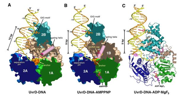
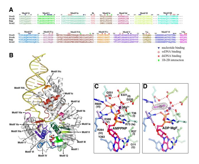
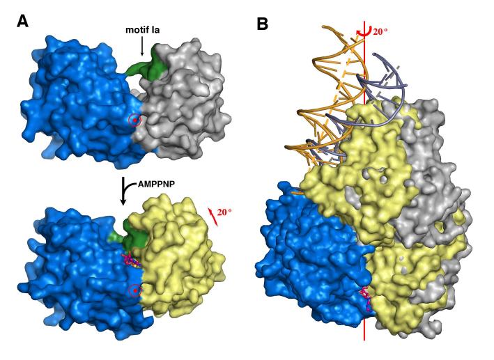
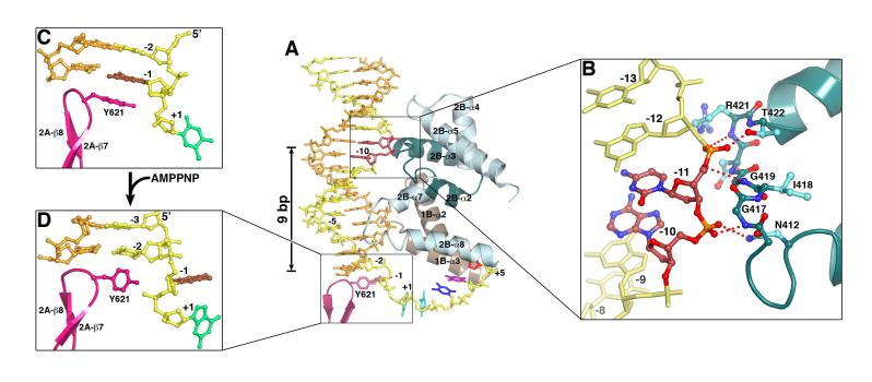
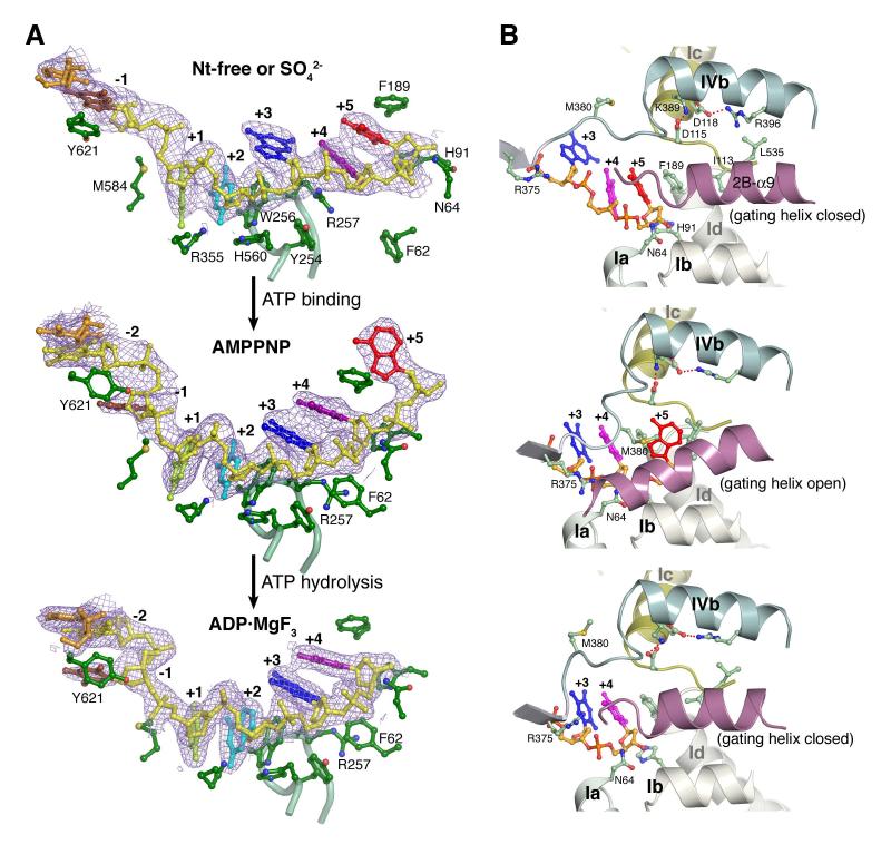
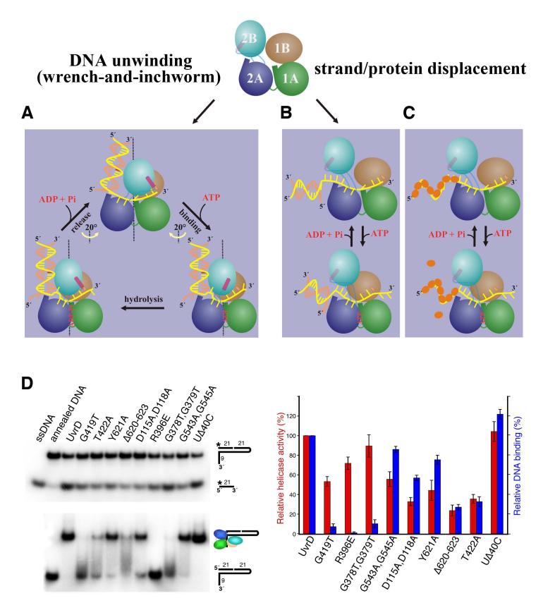
Helicases use the energy derived from nucleoside triphosphate hydrolysis to unwind double helices in essentially every metabolic pathway involving nucleic acids. Earlier crystal structures have suggested that DNA helicases translocate along a single-stranded DNA in an inchworm fashion. We report here a series of crystal structures of the UvrD helicase complexed with DNA and ATP hydrolysis intermediates. These structures reveal that ATP binding alone leads to unwinding of 1 base pair by directional rotation and translation of the DNA duplex, and ADP and Pi release leads to translocation of the developing single strand. Thus DNA unwinding is achieved by a two-part power stroke in a combined wrench-and-inchworm mechanism. The rotational angle and translational distance of DNA define the unwinding step to be 1 base pair per ATP hydrolyzed. Finally, a gateway for ssDNA translocation and an alternative strand-displacement mode may explain the varying step sizes reported previously.


Figures

Figure 1
Figure 1
Crystal structures of UvrD-DNA complexes. (A) binary complex, (B) AMPPNP and (c) ADP·MgF3 ternary complexes. Domain 1A, 1B, 2A and 2B are colored green, beige, blue and cyan and shown in molecular surface with the front of molecule removed to exposed the bound DNA in (A) and (B), and in ribbon diagram in (C). Functionally important regions are highlighted in orange (the GIG motif and anchor) and hot pink (the separation pin, ssDNA cap and gating helix). DNAs are shown as tubular models in (A) and (B) and stick models in (C). The translocated strand is colored yellow and the partner strand orange. The DNA base contacting the GIG motif is highlighted in red.
Figure 2
Figure 2
The conserved sequence motifs among UvrD, PcrA, Rep and Srs2. (A) Sequence alignment of the 8 ATPase motifs (in different shades of green (domain 1A) and blue and purple (2A), and 8 newly found DNA binding and domain interaction motifs (in brown to red colors). (B) The 16 motifs are mapped onto the UvrD-DNA-AMPPNP structure using the same color scheme as in (A). (C) and (D) Coordination of the AMPPNP and ADP·MgF3 by the 8 helicase motifs. Carbon atoms of the UvrD side chains are colored light green (domain 1A) and light blue and purple (2A). Nitrogen atoms are shown in blue, oxygen in red, and Mg2+ in dark purple. Water molecules are shown as red spheres. The Fo-Fc electron density of MgF3 is superimposed onto the model.
Figure 3
Figure 3
ATP-dependent domain rotation. (A) Domains 2A (blue) and 1A (grey or yellow) shown in molecular surface rotate towards each other by 20° upon binding of AMPPNP. For convenience, domain 2A is held stationary. Motif 1a is highlighted in green as a reference point. AMPPNP is shown as pink and orange ball-and-sticks. The view is down the rotation axis marked by the target sign. (B) An orthogonal view from (A) with the superimposed full UvrD-DNA binary (blue and silver) and ternary (blue, yellow and gold) complex structures. The DNA helical axis is shown as a grey (binary complex) or yellow (ternary complex) dashed line.
Figure 4
Figure 4
Interactions between UvrD and dsDNA. (A) The four HLH structures from domains 2B (light and dark cyan) and 1B (brown) interact with 14 to 16 bps. These interactions are similar between the binary and ternary complexes, while the ternary complex structure is shown. The separation pin (Y621) buttresses the end of DNA duplex. (B) A close-up view of the GIG motif and dsDNA interactions. (C) Stacking of Y621 with the-1 bp in the binary complex. (D) Unwinding of the-1 bp in the ternary complex, and the accompanying side chain conformational change of Y621.
Figure 5
Figure 5
Interactions between UvrD and ssDNA. (A) The interactions in the three distinct states of UvrD-DNA complexes. The backbone of -1 to +5 nts of the translocated strand is shown in yellow and the bases in distinct colors. A 2Fo-Fc electron density map corresponding to the ssDNA is shown in light purple. Carbon atoms of the protein side chains are shown in green, oxygen in red, nitrogen in blue and sulfur in yellow. The Cα trace of Motif III is shown as a light green tube. (B) Diagram of the gating helix conformation with the salt bridges at the domain 1B and 2B interface shown. Each corresponds to the nucleotide-binding state shown on the left.
Figure 6
Figure 6
Structural and functional duality of UvrD. (A) Cartoon presentations of the “wrench-and-inchworm mechanism” for DNA unwinding. Each domain is color-coded and the gating helix is highlighted in pink. (B) and (C) A mechanism for strand and protein displacement. The small orange ovals represent RecA-like proteins. (D) Assays of WT and mutant UvrDs in DNA unwinding (top left) and dsDNA binding (bottom left) using the nicked hairpin DNA substrate as diagramed. The relative helicase and DNA binding activities of each mutant UvrD are averaged from 3 measurements and plotted on the right in red (helicase) and blue (DNA binding).

References

    1. Abdel-Monem M, Chanal MC, Hoffmann-Berling H. DNA unwinding enzyme II of Escherichia coli. 1. Purification and characterization of the ATPase activity. Eur J Biochem. 1977;79:33–38. - PubMed
    1. Ali JA, Lohman TM. Kinetic measurement of the step size of DNA unwinding by Escherichia coli UvrD helicase. Science. 1997;275:377–380. - PubMed
    1. Arthur HM, Lloyd RG. Hyper-recombination in uvrD mutants of Escherichia coli K-12. Mol Gen Genet. 1980;180:185–191. - PubMed
    1. Becker PB, Horz W. ATP-dependent nucleosome remodeling. Annu Rev Biochem. 2002;71:247–273. - PubMed
    1. Bernstein DA, Zittel MC, Keck JL. High-resolution structure of the E.coli RecQ helicase catalytic core. Embo J. 2003;22:4910–4921. - PMC - PubMed

Publication types

MeSH terms