. 2006 Dec 29;127(7):1453-67.
doi: 10.1016/j.cell.2006.09.052.

Neuronal transcriptome of Aplysia: neuronal compartments and circuitry

Affiliations

Neuronal transcriptome of Aplysia: neuronal compartments and circuitry

Leonid L Moroz et al. Cell. .

Abstract

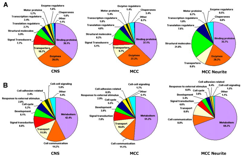
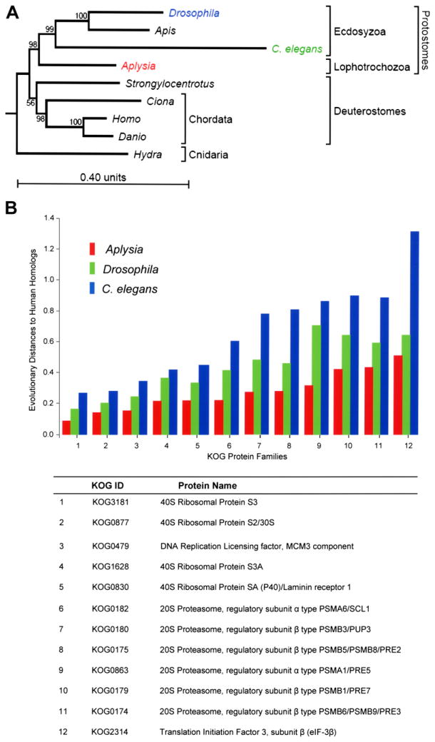
Molecular analyses of Aplysia, a well-established model organism for cellular and systems neural science, have been seriously handicapped by a lack of adequate genomic information. By sequencing cDNA libraries from the central nervous system (CNS), we have identified over 175,000 expressed sequence tags (ESTs), of which 19,814 are unique neuronal gene products and represent 50%-70% of the total Aplysia neuronal transcriptome. We have characterized the transcriptome at three levels: (1) the central nervous system, (2) the elementary components of a simple behavior: the gill-withdrawal reflex-by analyzing sensory, motor, and serotonergic modulatory neurons, and (3) processes of individual neurons. In addition to increasing the amount of available gene sequences of Aplysia by two orders of magnitude, this collection represents the largest database available for any member of the Lophotrochozoa and therefore provides additional insights into evolutionary strategies used by this highly successful diversified lineage, one of the three proposed superclades of bilateral animals.


Figures

Figure 1
Figure 1. Construction of an Aplysia Neurotranscriptomics Database
(A) Table summarizing statistics for the neuronal transcriptome project including collections of ESTs from identified MCC neuron and neuronal processes (neurites) of the same cell. (B) Aplysia californica, a free moving animal. Scale: 4 cm. (C) Schematic representation of the Aplysia central nervous system and its nine major ganglia, including paired buccal (BG), pedal (PeG), pleural (PlG), and cerebral (CG) as well as a single abdominal (AG) ganglion; a pair of neurosecretory clusters (Bag cells: Bc) is located in the abdominal ganglion and involved in the control of egg-laying behavior. (D) Dorsal view of the left and right cerebral ganglia of Aplysia and position of serotonergic modulatory neurons (MCC). All connective tissues were removed to reveal individual neurons located at the surface of the ganglia. Scale: 400 μm.
Figure 2
Figure 2. Comparison of ESTs Identified from CNS, MCC Somata, and MCC Neurites
(A and B) Annotated ESTs broken out according to major GO categories by both molecular function (A) and biological process (B) for ESTs obtained from the whole CNS, MCC somata, and MCC neurites. While the majority of categories stay relatively constant in each mRNA source, there are significantly fewer signal transducers and more structural molecules found in MCC neurites.
Figure 3
Figure 3. Aplysia in Evolutionary Context
(A) Phylogenetic relationship between Aplysia and other genomic model animals reconstructed based on the KOG (Tatusov et al., 2003) sequence analysis. The mollusc Aplysia californica (a representative of the Lophotrochozoa clade) is placed as a sister to the Arthropod (the fruit fly, Drosophila melanogaster, and the honey bee, Apis mellifera), and nematode (Caenorhabditis elegans) phyla representing the clade Ecdysozoa. The clade of Deuterostomes is represented here by chordates (Homo sapiens, the zebrafish Danio rerio, the ascidian Ciona intestinalis) and the sea urchin (Strongylocentrotus purpuratus). Therefore, this evolutionary analysis shows that Aplysia is more closely related to nematodes and insects than to vertebrates. The evolutionary distance (measured as branch length) from human to Aplysia is shorter, however, than the distance from human to Drosophila and C. elegans, suggesting that the amino acid replacement rate has been lower in the lineage leading to Aplysia than in the lineages leading to Drosophila and C. elegans. (B) Comparison of relative evolutionary distances for selected protein families between model organisms (C. elegans, Drosophila, Aplysia) and human homologs. The relative distances (the y axis) are measured for each KOG protein family (Tatusov et al., 2003) according to the JTT model of evolution (Jones et al., 1992). The distance units are expected amino acid replacements per site (for further details see Supplemental Data). Note that rates of evolutionary protein changes in the Aplysia lineage (red bars) were slower when compared to C. elegans or Drosophila lineages (blue and green bars).
Figure 4
Figure 4. Expression Patterns of Selected Neuronal Genes in the CNS of Aplysia californica (In Situ Hybridization)
RNA probes were designed based on cloned sequences to examine their expression in individual Aplysia ganglia. (A) Expression of serotonin transporter (AAK94482) in paired MCC neurons, in a dorsal view of the cerebral ganglion. (B) Novel isoform of pedal-type neuropeptide (DQ479396) is predominantly expressed in two symmetrical clusters of motoneurons (A/B groups) in the cerebral ganglion. (C) Expression of Huntingtin in the left pleural ganglion (LPl1 is the giant cholinergic neuron involved in control of mucus release from the body wall). (D) Acetylcholine Binding Protein (AAL37251) is exclusively expressed in glial type cell of Aplysia (the caudal part of the abdominal ganglion). (E) Expression of Achatin-like neuropeptide precursor (AY842441) in the abdominal ganglion. (F) Highly localized expression of Pleurin neuropeptide precursor (AY833131) in the right pleural ganglion. Scales: 300 μm (A and B); 200 μm (C); 100 μm (D); 350 μm (E); 120 μm (F).
Figure 5
Figure 5. Identification of L7 and Sensory Neuron-Specific Transcripts Using Microarrays
(A) Selected differentially expressed transcripts in L7 and sensory neurons. (B) Expression results from oligoarray experiments comparing L7 and sensory neurons. Spots representing transcripts common to the two neuronal types have been removed for clarity. (C–H) Neuron-specific expression (in situ hybridization) of selected transcripts identified in the microarray experiments (C, E, and H: abdominal ganglion; D and G: pleural ganglia; F: cerebral ganglia). Transcripts encoding Aplysia prothoraracicostatic peptide (PTSP) homolog (DQ375548, C and D), MIP-like neuropeptide (AAF80382, E) and Frizzled-2 (AY535406, H) are expressed in L7 (but not in sensory neurons, e.g., D), while BMP-like (AY485265, F) and TSH glycoprotein hormone β subunit-related protein (AAX35673), G) genes are expressed in sensory neurons. Note that MIP-like peptide prohormone and Frizzled-2 are also expressed in a subset of abdominal neurons, including some gill motoneurons and R2 neuron. Scales: 300 μm (C); 250 μm (D); 400 μm (E and H); 200 μm (F); 150 μm (G).

References

    1. Aguinaldo AM, Turbeville JM, Linford LS, Rivera MC, Garey JR, Raff RA, Lake JA. Evidence for a clade of nematodes, arthropods and other moulting animals. Nature. 1997;387:489–493. - PubMed
    1. Brannvall K, Hjelm H, Korhonen L, Lahtinen U, Lehesjoki AE, Lindholm D. Cystatin-B is expressed by neural stem cells and by differentiated neurons and astrocytes. Biochem Biophys Res Commun. 2003;308:369–374. - PubMed
    1. Brusca RC, Brusca GJ. Invertebrates. 2. Sunderland, MA: Sinauer Associates, Inc; 2003.
    1. Cash D, Carew TJ. A quantitative analysis of the development of the central nervous system in juvenile Aplysia californica. J Neurobiol. 1989;20:25–47. - PubMed
    1. Chen JY, Bottjer DJ, Oliveri P, Dornbos SQ, Gao F, Ruffins S, Chi H, Li CW, Davidson EH. Small bilaterian fossils from 40 to 55 million years before the Cambrian. Science. 2004;305:218–222. - PubMed

Publication types