. 2007 Jan 2;104(1):42-7.
doi: 10.1073/pnas.0609796104. Epub 2006 Dec 26.

Inhibitory complex of the transmembrane ammonia channel, AmtB, and the cytosolic regulatory protein, GlnK, at 1.96 A

Affiliations

Inhibitory complex of the transmembrane ammonia channel, AmtB, and the cytosolic regulatory protein, GlnK, at 1.96 A

Franz Gruswitz et al. Proc Natl Acad Sci U S A. .

Abstract

Ammonia conductance is highly regulated. A P(II) signal transduction protein, GlnK, is the final regulator of transmembrane ammonia conductance by the ammonia channel AmtB in Escherichia coli. The complex formed between AmtB and inhibitory GlnK at 1.96-A resolution shows that the trimeric channel is blocked directly by GlnK and how, in response to intracellular nitrogen status, the ability of GlnK to block the channel is regulated by uridylylation/deuridylylation at Y51. ATP and Mg(2+) augment the interaction of GlnK. The hydrolyzed product, adenosine 5'-diphosphate orients the surface of GlnK for AmtB blockade. 2-Oxoglutarate diminishes AmtB/GlnK association, and sites for 2-oxoglutarate are evaluated.


Conflict of interest statement

The authors declare no conflict of interest.

Figures

Fig. 1.
Fig. 1.
Physiological complex of AmtB and GlnK. (A) View from the cytoplasmic side down the threefold symmetry axis. (B) View within the plane of the membrane, 90° rotated about the horizontal with respect to A. The molecular surface of GlnK (red) and AmtB (blue) are displayed with different shades for each monomer of the two-trimer complex. Detergent molecules (yellow) are shown as sticks; atoms of partially ordered detergent are represented as spheres.
Fig. 2.
Fig. 2.
Monomer to monomer AmtB/Glnk interaction and comparison to uncomplexed structures. (A) Stereoview of a single inhibitory interaction (one asymmetric unit) between GlnK and AmtB. A ribbon diagram of AmtB (blue) and GlnK (red) is oriented identically to the matching colored monomers in Fig. 1. ADP bound to GlnK is shown in stick representation. (B) An overlay of the backbone of AmtB (blue) and GlnK (red) in the complex, compared with structures of the uncomplexed proteins [AmtB, 1U7G (black) and Amt1, 2B2H (green)]. Two loops of AmtB on the cytoplasmic side in the shaded region i are arranged differently from uncomplexed AmtB, with corresponding differences in 21 residues at the C terminus (dashed region ii). The complexed form of the AmtB C terminus is the same as that in uncomplexed Amt1. The T-loop of GlnK becomes highly ordered in the ADP-bound inhibitory complex vs. disordered in uncomplexed ATP-bound GlnK [2GNK (orange), dotted region iii].
Fig. 3.
Fig. 3.
Channel inhibition and ammonia conductance pathway. Blockade of the ammonia conduction pathway by R47 of GlnK (red) is in context (Center) and in detail (Left). The surface of AmtB (blue) is shown with residues 211–270 as a tubular backbone trace to reveal the hydrophobic channel behind (Right). Side chains of residues in AmtB along the conduction pathway (F107, F215,H168, and H318) and residues that interact with R47 (S263, D313, and carbonyl of C312) are in ball-and-stick representation with nitrogen (dark blue) and oxygen (light red) shown. The location of L259 (blue sphere on the tubular trace) and V299 that forms part of the surface is indicated. Ammonia positions (Am2, Am3, and Am4) seen only in uncomplexed AmtB are indicated as yellow spheres. In the complex, there are only two water/NH3 sites within the channel (Am6 and Am5). Waters within 6 Å of the channel and hydrophobic portals are shown as green spheres. A molecule of Tris (a quaternary ammonium cation, C+) displaced NH4+ at the periplasmic “recruitment site” (top right).
Fig. 4.
Fig. 4.
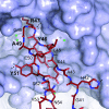
Specific interaction of the GlnK T-loop (red stick representation colored by atom) with AmtB (blue semitransparent surface and underlying sticks) is illustrated. The hydrogen bonds (black dashed lines) and geometry constitute a specific interface between channel and inhibitor. The sites that are subject to posttranslational modification in PII proteins at Y51 or at S49 in cyanobacteria are each found in pockets, such that when modified, they would displace the T-loop because of steric interference.
Fig. 5.
Fig. 5.
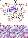
The ADP site between GlnK monomers. Residues are numbered with those of the second monomer labeled (†). (A) A cartoon of GlnK residues and bridging waters that support ADP. All dashed lines indicate hydrogen bonds with distances in the range 2.7–3.2 Å. (B) Stereoview of the electron density around the ADP site (σ-weighted 2FoFc map contoured at 1.0 σ). Labeled residues and colors are as in A.
Fig. 6.
Fig. 6.
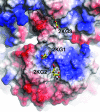
Docking of 2-oxoglutarate to pockets within GlnK (2KG1 and 2KG2) and in the interface with AmtB (2KG3). The electrostatic potential is shown on the solvent-accessible surface of the complex with a stick model of GlnK under the surface. 2-KG molecules (ball and stick, with carbons in yellow) as docked into the proposed 2-KG sites using DOCK6, are surrounded by positive electrostatic potential (blue). The 2KG1 and 2KG2 sites on GlnK are both adjacent to the T-loop and could account for disruption of the AmtB/GlnK complex by distorting the base of the T-loop. The proximity of the 2KG2 site to negatively charged ADP (ball and stick, carbons in black below the surface and directly behind the molecule in 2KG2) is consistent with the effect of Mg2+ as a counter ion.

References

    1. Broach J, Neumann C, Kustu S. J Bacteriol. 1976;128:86–98. - PMC - PubMed
    1. Knepper MA, Packer R, Good DW. Physiol Rev. 1989;69:179–249. - PubMed
    1. Saier MH, Jr, Eng BH, Fard S, Garg J, Haggerty DA, Hutchinson WJ, Jack DL, Lai EC, Liu HJ, Nusinew DP, et al. Biochim Biophys Acta. 1999;1422:1–56. - PubMed
    1. Thomas GH, Mullins JG, Merrick M. Mol Microbiol. 2000;37:331–344. - PubMed
    1. Monfort P, Kosenko E, Erceg S, Canales JJ, Felipo V. Neurochem Int. 2002;41:95–102. - PubMed

Publication types

MeSH terms

Associated data

LinkOut - more resources