. 2007 Jan 2;104(1):66-71.
doi: 10.1073/pnas.0609737104. Epub 2006 Dec 26.

Oxidized messenger RNA induces translation errors

Affiliations

Oxidized messenger RNA induces translation errors

Mikiei Tanaka et al. Proc Natl Acad Sci U S A. .

Abstract

To investigate the effect of RNA oxidation on normal cellular functions, we studied the translation of nonoxidized and oxidized luciferase mRNA in both rabbit reticulocyte lysate and human HEK293 cells. When HEK293 cells transfected with nonoxidized mRNA encoding the firefly luciferase protein were cultured in the presence of paraquat, there was a paraquat concentration-dependent increase in the formation of luciferase short polypeptides (SPs) concomitant with an increase in 8-oxoguanosine. Short polypeptides were also formed when the mRNA was oxidized in vitro by the Fe-ascorbate-H(2)O(2) metal-catalyzed oxidation system before its transfection into cells. Translation of the in vitro oxidized mRNA in rabbit reticulocyte lysate also led to formation of SPs. The SPs formed by either procedure contained the N-terminal and the C-terminal portions of the tagged luciferase. In addition, the oxidized mRNA was able to associate with ribosomes to form polysomes similar to those formed with nonoxidized mRNA preparations, indicating that the oxidized mRNAs are mostly intact; however, their translation fidelity was significantly reduced. Nevertheless, our results indicate that the SPs were derived from both premature termination of the translation process of the oxidized mRNA and the proteolytic degradation of the modified full-length luciferase resulting from translation errors induced by oxidized mRNA. In light of these findings, the physiological consequences of mRNA oxidation are discussed.


Conflict of interest statement

The authors declare no conflict of interest.

Figures

Fig. 1.
Fig. 1.
Detection of SPs in oxidatively stressed cells. HEK293 cells transfected with mRNA encoding both the FLAG- and c-Myc-tagged luciferase gene were cultured in the presence of paraquat for 6 h. (Upper) Immunoblots using anti-FLAG antibody (Left two gels) or anti-c-Myc antibody (Right two gels) were prepared from cells treated with or without MG132. (Lower) Immunoblots with anti-β-actin after stripping out the anti-FLAG and anti-c-Myc to verify equal loading.
Fig. 2.
Fig. 2.
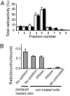
Oxidation of mRNA transfected to cells under oxidative stress. (A) Tritium-labeled mRNA extracted from transfected HEK293 cells treated with (solid bars) or without (open bars) 20 μM paraquat for 6 h was subjected to 1% agarose gel electrophoresis as described in Materials and Methods. The lane on a gel was excised into nine blocks from the top to the front end. The radioactivity in each block was measured. (B) Extracted mRNA (≈6,000 cpm) from transfected cells was immunoprecipitated with anti-8-oxoguanosine antibody. Where indicated, the mRNA was pretreated with RNase or DNase, or the antibody was pretreated with 8-oxoguanosine. The generation of 8-oxoguanosine was quantitated and expressed as the ratio of the bound radioactivity to that of the unbound form. Results were from three independent RNA samples.
Fig. 3.
Fig. 3.
Translation of oxidized mRNA in HEK293 cells. (A) Relative luciferase activity [F-luciferase/Renilla luciferase (R-luciferase)] observed with samples containing F-luciferase mRNA incubated for 1 h in the presence of the indicated amounts of a 1:1 mixture of ferrous iron and ascorbate, in the absence or presence of 1 mM EDTA before transfection as indicated. After transfection, cells were incubated for 12 h before measurement of luciferase activity. (B) Data obtained with F-luciferase mRNA incubated with 10 μM ascorbate (asc), 10 μM ferrous chloride (Fe), 10 μM ferrous chloride and ascorbate (Fe-asc), or no addition of ferrous iron and ascorbate (ctrl), under aerobic or anaerobic conditions before transfection.
Fig. 4.
Fig. 4.
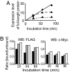
In vitro translation of oxidized mRNA. mRNA oxidized by iron–ascorbate and hydrogen peroxide was incubated in rabbit reticulocyte lysate supplemented with protease inhibitors as described in Materials and Methods. After incubation for the indicated times, the reaction mixture was subjected to Western blot analysis using anti-FLAG antibody or anti-c-Myc antibody. The Western blot intensity obtained with anti-FLAG and anti-c-Myc antibodies was normalized with the assumption that the full-length proteins contained both FLAG and c-Myc tags. (A) Time course for the relative amount of full-length protein formed with nonoxidized mRNA (♦), mRNA oxidized for 1 h with 5 μM 1:1 iron–ascorbate and 1 μM H2O2 (■), or mRNA oxidized for 1 h with only 5 μM H2O2 (▴). (B) Ratio of the SPs to total protein/peptide generated, as monitored by Western blotting (WB) with anti-FLAG (N-terminal) (Left) or anti-c-Myc (C-terminal) (Right) antibodies. Solid black bar, gray bar, and open bar represent data for nonoxidized mRNA, mRNA oxidized with 1 μM H2O2, or mRNA oxidized with 5 μM H2O2, respectively, as described above.
Fig. 5.
Fig. 5.
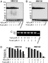
Generation of SPs by translation of oxidized mRNA. (A and B) (Upper) HEK293 cells were transfected with in vitro oxidized mRNA, mock mRNA (mRNA unrelated to luciferase), or truncated mRNA and incubated with (A) or without (B) MG132. The translation products were subjected to Western blot analysis using the anti-FLAG antibody. (Lower) Anti-β-actin immunoblots to verify equal loading for each lane. (C) The mRNA samples were examined by agarose gel electrophoresis followed by ethidium bromide staining. (D) The expression levels of the full-length proteins, quantified by using a fluorescence-based assay (black bars) together with luciferase activity (gray bars) in cell lysates, were compared in cells treated either with (Left) or without (Right) MG132.
Fig. 6.
Fig. 6.
Binding of oxidized mRNA to polysomes. (A) In vitro oxidized 3H-labeled RNA was transfected into HEK293 cells. The cell lysate was subjected to sucrose density centrifugation and fractionated as described in Materials and Methods. The radioactivity was measured for each fraction. (B) Percent recovery of the oxidized mRNA in the polysome fraction. (C) Relative luciferase activity in the cell lysate.

References

    1. Stadtman ER, Berlett BS. Chem Res Toxicol. 1997;10:485–494. - PubMed
    1. Beckman KB, Ames BN. Physiol Rev. 1998;78:547–584. - PubMed
    1. Evans MD, Dizdaroglu M, Cooke MS. Mutat Res. 2004;567:1–61. - PubMed
    1. Hofer T, Badouard C, Bajak E, Ravanat JL, Mattsson A, Cotgreave IA. Biol Chem. 2005;386:333–337. - PubMed
    1. Fiala ES, Conaway CC, Mathis JE. Cancer Res. 1989;49:5518–5522. - PubMed

Publication types

LinkOut - more resources