. 2007 Jan 2;104(1):335-40.
doi: 10.1073/pnas.0607450104. Epub 2006 Dec 26.

Activity-dependent neurotransmitter-receptor matching at the neuromuscular junction

Affiliations

Activity-dependent neurotransmitter-receptor matching at the neuromuscular junction

Laura N Borodinsky et al. Proc Natl Acad Sci U S A. .

Abstract

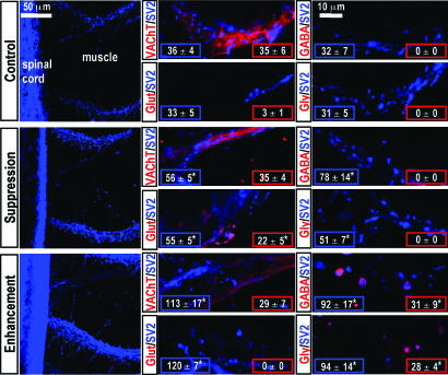
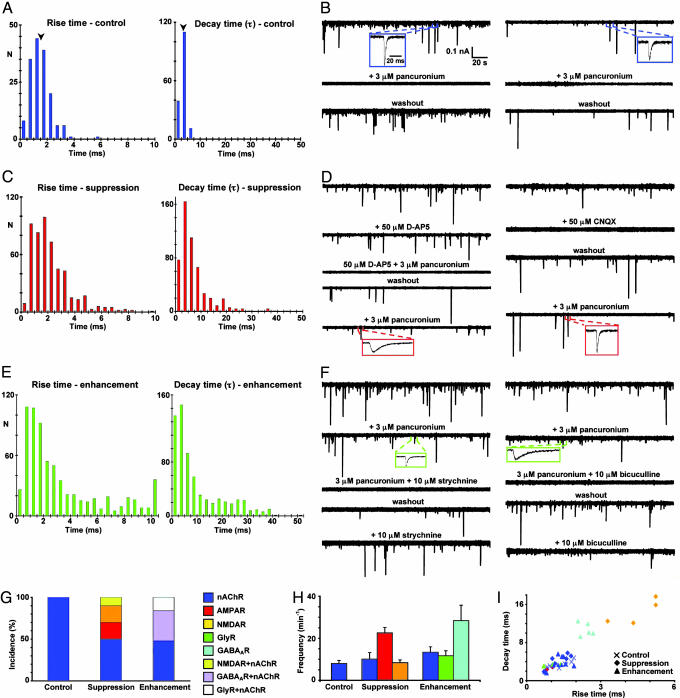
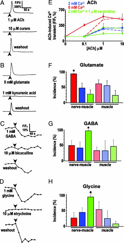
Signaling in the nervous system requires matching of neurotransmitter receptors with cognate neurotransmitters at synapses. The vertebrate neuromuscular junction is the best studied cholinergic synapse, but the mechanisms by which acetylcholine is matched with acetylcholine receptors are not fully understood. Because alterations in neuronal calcium spike activity alter transmitter specification in embryonic spinal neurons, we hypothesized that receptor expression in postsynaptic cells follows changes in transmitter expression to achieve this specific match. We find that embryonic vertebrate striated muscle cells normally express receptors for glutamate, GABA, and glycine as well as for acetylcholine. As maturation progresses, acetylcholine receptor expression prevails. Receptor selection is altered when early neuronal calcium-dependent activity is perturbed, and remaining receptor populations parallel changes in transmitter phenotype. In these cases, glutamatergic, GABAergic, and glycinergic synaptic currents are recorded from muscle cells, demonstrating that activity regulates matching of transmitters and their receptors in the assembly of functional synapses.


Conflict of interest statement

The authors declare no conflict of interest.

Figures

Fig. 1.
Fig. 1.
Expression of nAChR, NMDAR, AMPAR, GABAAR, and GlyR transcripts and protein in skeletal muscle during normal development and after alterations in neuronal activity. (A) RT-PCR was used for detection of subunit transcripts of five neurotransmitter receptors in muscle, notochord, and neural tube at three stages of development. Tissue-specific RNA was analyzed from embryos at 1 day (stage 22, Top), at 1.3 days (stage 28, Middle) and from larvae at 3 days (stage 40, Bottom). Primers were designed from predicted Xenopus sequences for nAChRα1, NR1, GluR1, GABAARβ2, and GlyRα1 subunits and for neuronal markers NeuroD and neurogenin-related-protein-1 (NGNR-1). (B and C) Multiple classes of transmitter receptors are expressed in embryonic skeletal muscle in vivo. Whole mounts from 1.3-day (stage 28) embryos and 3-day (stage 40) larvae were labeled for myosin and nuclei (B), nAChR, NMDAR, AMPAR, GABAAR, and GlyR (C) with probes noted above each column. Images of chevrons of mononucleate muscle cells are representative Z series projections obtained from confocal stacks of 20 optical sections of 62,500 μm2 area. (C Insets) Percent of labeled volume. Values are mean ± SEM, n ≥ 5 embryos for each probe. ∗, P < 0.001 when compared with stage 40 for each probe. (D) Alterations of neuronal Ca2+ spike activity change in vivo expression of transmitter receptors in larval skeletal muscle. Whole mounts from activity-manipulated 3-day (stage 40) larvae were labeled for transmitter receptors as in C. Manipulation of activity was achieved by implanting beads impregnated with 30 μM tetrodotoxin, 200 nM calcicludine, 10 μM GVIA ω-conotoxin, and 10 μM flunarizine (Upper, Ca2+ spike activity suppression) or with 1 mM veratridine (Lower, Ca2+ spike activity enhancement). Specimens were stained and labeling was quantified (Insets) as in C. Values are mean ± SEM for n ≥ 5 embryos for each probe. ∗, P < 0.001 when compared with stage 40 control for each probe.
Fig. 2.
Fig. 2.
Activity-dependent neurotransmitter expression in presynaptic terminals in the axial musculature. Whole mounts from control and activity-manipulated 3-day (stage 40) larvae were immunolabeled for a presynaptic marker, SV2 (in blue), and for transmitter phenotype (in red): VAChT (cholinergic), glutamate (glutamatergic), GABA (GABAergic), and glycine (glycinergic). (Left) Low magnification images are representative Z series projections obtained from confocal stacks of 50 optical sections of 44,100 μm2 area of muscle and spinal cord. (Center and Right) High magnification images are representative Z series projections of five optical sections of muscle from the top of the same stacks. n = 5 embryos for each transmitter phenotype. (Insets) Numbers of synaptic puncta labeled for SV2 and for transmitter per 33,443 μm2 area of muscle. Values are mean ± SEM for n = 5 single optical sections per probe from five embryos. ∗, P < 0.05 compared with control.
Fig. 3.
Fig. 3.
Matching of neurotransmitters and receptors at neuromuscular junctions in vivo. Whole-cell recordings from muscle cells of the axial musculature of 3-day (stage 40) control (A and B), Ca2+ spike-suppressed (C and D), and Ca2+ spike-enhanced (E and F) larvae were performed in the presence of 2 mM Ca2+, Mg2+-free saline, and 3 μM TTX; Vh = −80 mV. (A, C, and E) Rise and decay time distributions for mpscs, including only mpscs with decay times fit by single exponentials. N, number of mpscs (≥6 embryos for each group). Arrowheads in A indicate mean values. (B, D, and F) Examples of pharmacological blockade of mpscs from two muscle cells in each group of embryos. Single mpscs are shown on an expanded time base to illustrate their kinetics. (G) Incidence of cholinergic, glutamatergic, GABAergic, and glycinergic NMJs. Bars represent the percent of each class of NMJ in each group based on blockade by different receptor antagonists. Bars represented by a single receptor are derived from NMJs in which mpscs were blocked completely by a single receptor antagonist. Bars corresponding to two receptors are derived from NMJs in which mpscs were blocked completely only by the combination of two receptor antagonists. (H) Frequencies of pharmacologically isolated mpscs in each group. Values are mean ± SEM. (I) Rise and decay times for pharmacologically identified mpscs. ×, control; ◇, spike-suppressed; ▵, spike-enhanced. Each point is the mean rise time and decay time for mpscs recorded from a single muscle cell. (GI) Values are from 6 control, 12 spike-suppressed, and 13 spike-enhanced NMJs.
Fig. 4.
Fig. 4.
Sensitivity of uninnervated muscle cells to ACh, glutamate, GABA, and glycine depends on neuronal Ca2+ spike activity in vitro. Muscle cells cocultured with neurons (nerve-muscle) and muscle cells cultured alone (muscle) for 18–24 h were loaded with a fluorescent Ca2+ indicator or Cl sensor dye to image responses to ACh and glutamate or GABA and glycine, respectively. Imaging was performed in 2 mM Ca2+ culture medium. Glutamate responses were recorded in the presence of 10 μM curare or 3 μM pancuronium to exclude indirect nAChR-mediated responses. (AD) Traces are responses of uninnervated single muscle cells to neurotransmitters applied at times indicated by arrowheads. (EH) Analysis of the incidence of responses to transmitters under different culture conditions. ∗, P < 0.01 when compared with the 2 mM Ca2+ condition for nerve-muscle cultures. (E) Solid lines represent nerve-muscle cultures, and dashed lines represent muscle cultures. Values are mean ± SEM from >40 cells from five independent cultures for each transmitter.

Similar articles

Cited by

References

    1. Borodinsky LN, Root CM, Cronin JA, Sann SB, Gu X, Spitzer NC. Nature. 2004;429:523–530. - PubMed
    1. Brosenitsch TA, Katz DM. Mol Cell Neurosci. 2002;20:447–457. - PubMed
    1. Gomez-Lira G, Lamas M, Romo-Parra H, Gutierrez R. J Neurosci. 2005;25:6939–6946. - PMC - PubMed
    1. Shi J, Townsend M, Constantine-Paton M. Neuron. 2000;28:103–114. - PubMed
    1. Catalano SM, Chang CK, Shatz CJ. J Neurosci. 1997;17:8376–8390. - PMC - PubMed

Publication types

MeSH terms