. 2007 Jan 9;104(2):582-7.
doi: 10.1073/pnas.0606699104. Epub 2006 Dec 26.

Regulation of innate antiviral defenses through a shared repressor domain in RIG-I and LGP2

Affiliations

Regulation of innate antiviral defenses through a shared repressor domain in RIG-I and LGP2

Takeshi Saito et al. Proc Natl Acad Sci U S A. .

Abstract

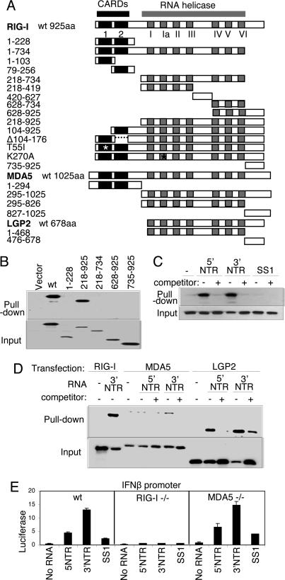
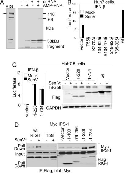
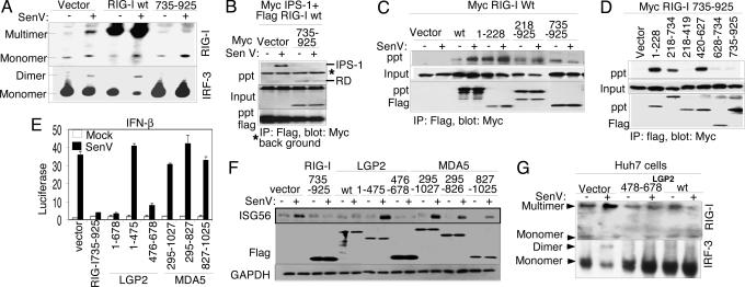
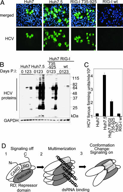
RIG-I is an RNA helicase containing caspase activation and recruitment domains (CARDs). RNA binding and signaling by RIG-I are implicated in pathogen recognition and triggering of IFN-alpha/beta immune defenses that impact cell permissiveness for hepatitis C virus (HCV). Here we evaluated the processes that control RIG-I signaling. RNA binding studies and analysis of cells lacking RIG-I, or the related MDA5 protein, demonstrated that RIG-I, but not MDA5, efficiently binds to secondary structured HCV RNA to confer induction of IFN-beta expression. We also found that LGP2, a helicase related to RIG-I and MDA5 but lacking CARDs and functioning as a negative regulator of host defense, binds HCV RNA. In resting cells, RIG-I is maintained as a monomer in an autoinhibited state, but during virus infection and RNA binding it undergoes a conformation shift that promotes self-association and CARD interactions with the IPS-1 adaptor protein to signal IFN regulatory factor 3- and NF-kappaB-responsive genes. This reaction is governed by an internal repressor domain (RD) that controls RIG-I multimerization and IPS-1 interaction. Deletion of the RIG-I RD resulted in constitutive signaling to the IFN-beta promoter, whereas RD expression alone prevented signaling and increased cellular permissiveness to HCV. We identified an analogous RD within LGP2 that interacts in trans with RIG-I to ablate self-association and signaling. Thus, RIG-I is a cytoplasmic sensor of HCV and is governed by RD interactions that are shared with LGP2 as an on/off switch controlling innate defenses. Modulation of RIG-I/LGP2 interaction dynamics may have therapeutic implications for immune regulation.


Conflict of interest statement

The authors declare no conflict of interest.

Figures

Fig. 1.
Fig. 1.
Constructs and RNA binding properties. (A) Domain structure of the RIG-I, MDA5, and LGP2 constructs showing the positions of the tandem CARDs and the RNA helicase domain and its subdomains conserved among the helicase superfamily (15). Point mutations are indicated by an asterisk. The amino acid region encoded by each construct is shown at left. (B) Extract from Huh 7 cells that were transfected with empty vector or plasmid expressing Flag-tagged RIG-I constructs encoding WT (wt) RIG-I or the indicated amino acid regions of RIG-I were mixed with pIC-agarose beads and subjected to pull-down assay for dsRNA binding. RIG-I proteins within the input material (Lower) and recovered as pull-down product (Upper) were evaluated by immunoblotting using anti-Flag antibody. (C and D) Cytoplasmic fraction (20 μg) from 293 cells transfected with plasmid encoding Flag-tagged WT RIG-I (C) or with Flag RIG-I wt, Flag-MDA5 wt, or Flag-LGP2 wt (D) were mixed with 1 μg of in vitro-transcribed biotin-UTP HCV 5′ NTR RNA, 3′ NTR RNA, or SS1 RNA alone (−) or with an excess of unlabeled homologous competitor RNA (+). RNA–protein complexes were recovered by pull-down assay using streptavidin affinity gel. Flag-tagged protein within the pull-down fraction or 25% of input material was analyzed by immunoblotting using anti-Flag antibody. (E) wt, RIG-I-null, or MDA5-null MEFs were cotransfected with plasmids encoding constitutive Renilla luciferase and firefly luciferase controlled by the murine IFN-β promoter (2). After 24 h, the cells were mock-transfected or transfected with 10 ng of in vitro-transcribed, gel-purified RNA encoding the HCV 5′ NTR, 3′ NTR, or SS1 region (6). After 24 h, the cells were harvested, and extracts were subjected to dual luciferase assay. Bars show relative luciferase values and SD.
Fig. 2.
Fig. 2.
An RD controls RIG-I signaling. (A) Recombinant RIG-I protein was digested with trypsin in the presence or absence of dsRNA and AMP-PNP as indicated. After termination of the reaction, the mixtures were analyzed by immunoblotting using anti-RIG-I mAb, which reacts with the C-terminal portion of RIG-I. Bars indicate the undigested 115-kDa RIG-I input protein (Left) and a 30-kDa protected digestion fragment (Right). (B) Huh 7 cells were cotransfected with luciferase promoter constructs and the indicated RIG-I expression constructs. After 24 h, the cells were mock-infected or infected with SenV. Cells were harvested 20 h later for dual luciferase assay. Bars show the mean relative IFN-β promoter–luciferase levels (± SD). (C) Huh 7.5 cells were cotransfected with promoter luciferase plasmids and empty vector or the indicated RIG-I expression plasmids. Cells were mock- or SenV-infected as indicated and were processed as above for dual luciferase assay (Left) or were harvested and analyzed by immunoblot assay for ISG56, Flag-RIG-I construct (Flag), and GAPDH abundance as shown (Right). (D) Huh7.5 cells were cotransfected with plasmids encoding Myc-IPS-1 and vector control or the indicated RIG-I constructs. After 16 h, the cells were mock-infected or infected with SenV as shown, cultured for 16 h, and harvested for anti-Flag immunoprecipitation (IP) and immunoblot assays as described in ref. . Shown are Flag-RIG-I protein abundance in the input material (Bottom) and Myc-IPS within the IP product (Top) or input material (Middle).
Fig. 3.
Fig. 3.
Mechanism of regulation by the RIG-I RD. Where indicated, cells were mock-infected or infected with SenV for 16 h before harvesting. (A) Stable Huh7 cell lines expressing vector alone, RIG-I wt, or RIG-I 735–925 were mock-infected or infected with SenV, and protein extracts were subjected to native PAGE and immunoblot analysis with anti-RIG-I antibody (Upper) or anti-IRF-3 antibody (Lower). Dimer/multimer and monomer protein forms are indicated. (B–D) Huh7 cells were cotransfected with plasmids encoding Myc-IPS-1, Flag-RIG-I wt, and vector or Myc-RIG-I 735–925 (B); Myc-RIG-I wt and the indicated Flag construct (C); or Myc-RIG-I 735–925 and the indicated Flag construct (D). Cells were infected as shown and harvested, and extracts were analyzed by immunoprecipitation (IP) and immunoblot assays. Shown are the abundance of Myc-tagged protein within anti-Flag IP products (Top), input Myc-tagged protein (Middle), and input Flag-tagged protein (Bottom). (E and F) Huh 7 cells were transfected with Renilla luciferase, IFN-β-luciferase plasmids, and plasmids encoding vector alone or the indicated Flag-tagged RIG-I, LGP2, or MDA5 constructs. After SenV infection, the cells were harvested and extracts were subjected to dual luciferase assay (E) (bars show relative luciferase and SD) and to immunoblot assay for abundance of ISG56, Flag-tagged protein (Flag), and GAPDH (F). (G) Anti-RIG-I (Upper) or anti-IRF-3 (Lower) immunoblot of Huh7 cell extracts separated by native PAGE. Protein monomer and multimer/dimer forms are indicated. Cells were transfected with vector control or expression plasmid encoding Flag-LGP2 478–378 or Flag-LGP2 wt. Extracts were prepared after SenV or mock infection.
Fig. 4.
Fig. 4.
The RD regulates cell permissiveness to HCV. Huh7, Huh7.5, Huh RIG-I wt, or Huh7-RIG-I-735–925 cells were infected with JFH-1 HCV 2A at a multiplicity of infection of 0.5. (A) After 48 h, cells were immunostained with HCV 2A antiserum (green). Nuclei were visualized by staining the cells with DAPI (blue). (B) Anti-HCV (Upper) and GAPDH immunoblot (Lower) of extracts from mock-infected cells (0) or from cells infected with JFH1 for 1, 2, or 3 days as indicated. The positions of HCV proteins are shown and have been defined previously (10). (C) Titer of HCV in supernatants collected from the indicated cell cultures 48 h after infection. (D) Model of RIG-I autoregulation and activation by virus infection. The RIG-I CARDs and domains encoding the helicase region and the RD are indicated.

References

    1. Meylan E, Tschopp J, Karin M. Nature. 2006;442:39–44. - PubMed
    1. Yoneyama M, Kikuchi M, Natsukawa T, Shinobu N, Imaizumi T, Miyagishi M, Taira K, Akira S, Fujita T. Nat Immunol. 2004;5:730–737. - PubMed
    1. Kang DC, Gopalkrishnan RV, Wu Q, Jankowsky E, Pyle AM, Fisher PB. Proc Natl Acad Sci USA. 2002;99:637–642. - PMC - PubMed
    1. Yoneyama M, Kikuchi M, Matsumoto K, Imaizumi T, Miyagishi M, Taira K, Foy E, Loo YM, Gale M, Jr, Akira S, et al. J Immunol. 2005;175:2851–2858. - PubMed
    1. Kato H, Takeuchi O, Sato S, Yoneyama M, Yamamoto M, Matsui K, Uematsu S, Jung A, Kawai T, Ishii KJ, et al. Nature. 2006;441:101–105. - PubMed

Publication types