

Par4 is required for platelet thrombus propagation but not fibrin generation in a mouse model of thrombosis
- PMID: 17190826
- PMCID: PMC1765451
- DOI: 10.1073/pnas.0610188104
Par4 is required for platelet thrombus propagation but not fibrin generation in a mouse model of thrombosis
Abstract
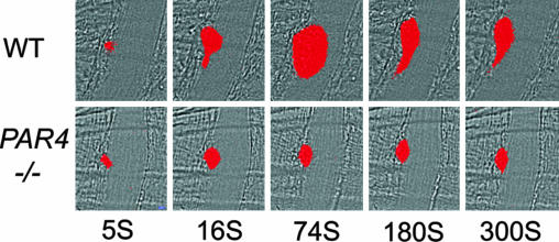
Thrombin, a central mediator of hemostasis and thrombosis, converts fibrinogen to fibrin and is a potent platelet activator. Activated platelets provide a surface for assembly of the tenase and prothrombinase complexes required for thrombin generation. The role of thrombin-induced platelet activation in platelet accumulation and its interplay with fibrin deposition during thrombus assembly has not been fully defined. We studied these processes during laser-induced thrombus formation by using real-time digital fluorescence microscopy in mice lacking protease-activated receptor-4 (Par4), which is necessary for thrombin responsiveness in mouse platelets. Juxtamural platelet accumulation immediately after laser injury was not different in wild-type and Par4(-/-) mice. However, subsequent growth of platelet thrombi was markedly diminished in Par4(-/-) mice. At the time of maximal thrombus size in wild type, platelet accumulation was more than 10-fold higher in wild type than in Par4(-/-) mice. P-selectin expression, a marker of platelet activation, was reduced and delayed in Par4(-/-) thrombi. In contrast to platelet activation and accumulation, the rate and amount of fibrin deposition, predominantly intramural and juxtamural in this model, were indistinguishable in Par4(-/-) and wild-type mice. These results suggest that platelet activation by thrombin is necessary for normal propagation of a platelet thrombus at a distance from the injured vessel wall and hence for normal thrombus growth. However, platelet activation by thrombin is unnecessary for initial and limited accumulation of platelets at or near the vessel wall, and this limited accumulation of platelets and/or platelet-independent mechanism(s) of thrombin generation are sufficient for normal fibrin deposition in this model.
Conflict of interest statement
The authors declare no conflict of interest.
Figures


Similar articles
-
Factor VIII contributes to platelet-fibrin thrombus formation via thrombin generation under low shear conditions.Thromb Res. 2009 Nov;124(5):601-7. doi: 10.1016/j.thromres.2009.06.035. Epub 2009 Aug 6. Thromb Res. 2009. PMID: 19660789
-
Thrombin-initiated platelet activation in vivo is vWF independent during thrombus formation in a laser injury model.J Clin Invest. 2007 Apr;117(4):953-60. doi: 10.1172/JCI30537. Epub 2007 Mar 22. J Clin Invest. 2007. PMID: 17380206 Free PMC article.
-
Platelets possess and require an active protein palmitoylation pathway for agonist-mediated activation and in vivo thrombus formation.Arterioscler Thromb Vasc Biol. 2007 Jun;27(6):1478-85. doi: 10.1161/ATVBAHA.106.139287. Epub 2007 Feb 15. Arterioscler Thromb Vasc Biol. 2007. PMID: 17303775
-
Role of platelet P-selectin and microparticle PSGL-1 in thrombus formation.Trends Mol Med. 2004 Apr;10(4):171-8. doi: 10.1016/j.molmed.2004.02.008. Trends Mol Med. 2004. PMID: 15059608 Review.
-
The interaction of thrombin with blood platelets.Platelets. 2005 Nov;16(7):373-85. doi: 10.1080/09537100500123568. Platelets. 2005. PMID: 16236598 Review.
Cited by
-
Genetic deletion of platelet PAR4 results in reduced thrombosis and impaired hemostatic plug stability.J Thromb Haemost. 2022 Feb;20(2):422-433. doi: 10.1111/jth.15569. Epub 2021 Nov 10. J Thromb Haemost. 2022. PMID: 34689407 Free PMC article.
-
Substituted indoles as selective protease activated receptor 4 (PAR-4) antagonists: Discovery and SAR of ML354.Bioorg Med Chem Lett. 2014 Oct 1;24(19):4708-4713. doi: 10.1016/j.bmcl.2014.08.021. Epub 2014 Aug 15. Bioorg Med Chem Lett. 2014. PMID: 25176330 Free PMC article.
-
DNAse-dependent, NET-independent pathway of thrombus formation in vivo.Proc Natl Acad Sci U S A. 2021 Jul 13;118(28):e2100561118. doi: 10.1073/pnas.2100561118. Proc Natl Acad Sci U S A. 2021. PMID: 34260389 Free PMC article.
-
Protease-activated receptor 4: from structure to function and back again.Br J Pharmacol. 2016 Oct;173(20):2952-65. doi: 10.1111/bph.13455. Epub 2016 Mar 10. Br J Pharmacol. 2016. PMID: 26844674 Free PMC article. Review.
-
Relipidated tissue factor linked to collagen surfaces potentiates platelet adhesion and fibrin formation in a microfluidic model of vessel injury.Bioconjug Chem. 2011 Oct 19;22(10):2104-9. doi: 10.1021/bc200326v. Epub 2011 Sep 8. Bioconjug Chem. 2011. PMID: 21902184 Free PMC article.
References
-
- Coughlin SR. Nature. 2000;407:258–264. - PubMed
-
- Sambrano GR, Weiss EJ, Zheng YW, Huang W, Coughlin SR. Nature. 2001;413:74–78. - PubMed
-
- Weiss EJ, Hamilton JR, Lease KE, Coughlin SR. Blood. 2002;100:3240–3244. - PubMed
-
- Hamilton JR, Cornelissen I, Coughlin SR. J Thromb Haemostasis. 2004;2:1429–1435. - PubMed
Publication types
MeSH terms
Substances
LinkOut - more resources
Full Text Sources
Other Literature Sources
Medical
Molecular Biology Databases
