. 2007 Jan 22;204(1):93-103.
doi: 10.1084/jem.20061598. Epub 2006 Dec 26.

An essential role for RasGRP1 in mast cell function and IgE-mediated allergic response

Affiliations

An essential role for RasGRP1 in mast cell function and IgE-mediated allergic response

Yan Liu et al. J Exp Med. .

Abstract

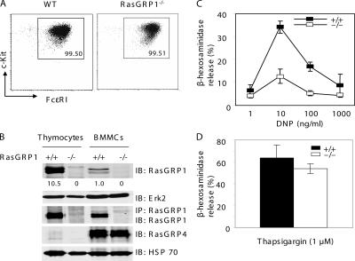
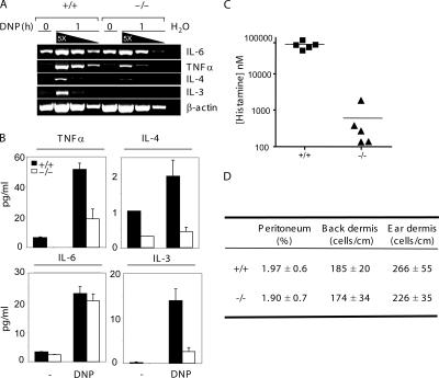
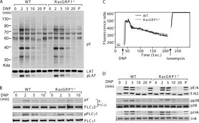
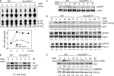
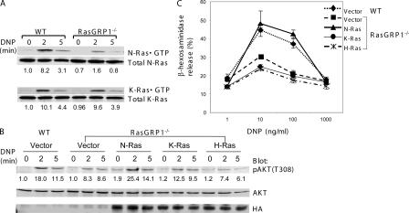
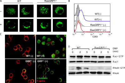
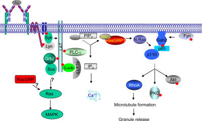
Cross-linking of the FcepsilonRI activates the phosphatidyl inositol 3 kinase (PI3K) and mitogen-activated protein kinase pathways. Previous studies demonstrate that Ras guanyl nucleotide-releasing protein (RasGRP)1 is essential in T cell receptor-mediated Ras-Erk activation. Here, we report that RasGRP1 plays an important role in FcepsilonRI-mediated PI3K activation and mast cell function. RasGRP1-deficient mice failed to mount anaphylactic allergic reactions. RasGRP1-/- mast cells had markedly reduced degranulation and cytokine production. Although FcepsilonRI-mediated Erk activation was normal, PI3K activation was diminished. Consequently, activation of Akt, PIP3-dependent kinase, and protein kinase C delta was defective. Expression of a constitutively active form of N-Ras could rescue the degranulation defect and Akt activation. We further demonstrated that RasGRP1-/- mast cells were defective in granule translocation, microtubule formation, and RhoA activation. Our results identified RasGRP1 as an essential regulator of mast cell function.


Figures

Figure 1.
Figure 1.
RasGRP1 in FcɛRI-mediated degranulation. (A) Expression of c-Kit and FcɛRI in WT and RasGRP1−/− BMMCs. (B) Expression of RasGRP1 and RasGRP4 in mast cells. Mast cell lysates were analyzed by Western blotting with anti-RasGRP1, RasGRP4, Erk2, or HSP70 antibodies or immunoprecipitated with anti-RasGRP1 antibodies. The numbers shown are relative quantification of RasGRP1 in thymocytes and BMMCs. (C) Effect of RasGRP1 inactivation on FcɛRI-mediated degranulation. BMMCs were sensitized with anti-DNP IgE and then stimulated with various concentrations of DNP-HSA for 10 min. Degranulation data were expressed as percentages of the released verses total β-hexosaminidase activity and reflect three independent experiments. (D) Thapsigargin-induced degranulation.
Figure 2.
Figure 2.
Effect of RasGRP1 deficiency on cytokine release and passive systemic anaphylaxis. (A) FcɛRI-induced cytokine RNA synthesis in WT and RasGRP1−/− BMMCs. Sensitized BMMCs were stimulated with 30 ng/ml DNP-HSA for 1 h or left unstimulated before RNA preparation. cDNAs were serially diluted and used in RT-PCR. Data shown are representative of two independent experiments with similar results. (B) FcɛRI-induced cytokine secretion. Data shown are representative of two independent experiments with similar results. (C) Effect of RasGRP1 inactivation on passive systemic anaphylaxis. WT and RasGRP1−/− mice were sensitized with anti-DNP IgE. Anaphylaxis was induced by injection with DNP-HSA. Histamine concentration in the blood was determined by ELISA (n = 5 for both WT and RasGRP1−/− mice). The horizontal bars indicate mean values. (D) Normal numbers of mast cells in the peritoneum, back skin, and ear dermis.
Figure 3.
Figure 3.
FcɛRI-mediated proximal signaling in WT and RasGRP1−/− BMMCs. (A) Tyrosine phosphorylation of proteins after FcɛRI engagement. After sensitization with anti-DNP IgE, BMMCs were stimulated with DNA-HSA or PMA (P) for 5 min. Total lysates were blotted with an anti-phosphotyrosine (pY) or anti-LATpY191 antibody. A similar amount of lysates loaded in each lane was indicated by an anti-LAT blot. (B) Tyrosine phosphorylation of PLCγ1 and PLCγ2. (C) FcɛRI-induced calcium influx. Sensitized mast cells were loaded with Indo-1 and stimulated with DNP-HSA. The fluorescence emission ratio at 405–495 nm was monitored by flow cytometry. (D) Activation of MAPKs. Whole cell lysates were analyzed by Western blotting with antibodies against Erk, Jnk, and p38 and their phosphorylated forms.
Figure 4.
Figure 4.
Impaired PI3K pathway in RasGRP1−/− BMMCs. (A) Effect of RasGRP1 deficiency on FcɛRI-induced PIP3 production in RasGRP1−/− BMMCs. Sensitized cells were labeled with [32P]orthophosphate and stimulated with DNP-HSA. Lipids were then extracted and subjected to thin layer chromatography. Increased PIP3 production is plotted in the bottom panel. Data shown were from a representative of three independent experiments. (B) Gab2 phosphorylation and its association with p85. Lysates were immunoprecipitated with anti-Gab2 followed by Western blotting with anti-pY, Gab2, and p85 antibodies. (C) FcɛRI-induced SHIP1 (Tyr1020) phosphorylation. Whole cell lysates were blotted with anti-pSHIP (Tyr1020) and SHIP antibodies. (D and E) Effect of RasGRP1 deficiency on the PI3K pathway. DNP-HSA or PMA (P) -activated mast cell lysates were analyzed by Western blotting with antibodies against phosphorylated AKT (Ser473 and Thr308), PDK1 (Ser241), PKCδ (Thr505), and GSK3β (Ser9). The numbers shown were normalized relative intensities for the phosphorylated Akt, PKD1, and PKCδ, respectively.
Figure 5.
Figure 5.
Activation of N-Ras by RasGRP1. (A) Reduced activation of N-Ras in RasGRP1−/− BMMCs. Sensitized WT and RasGRP1−/− cells were activated with DNP-HSA. Lysates were subjected to GST-Raf-RBD precipitation followed by Western blotting with anti–N-Ras and anti–K-Ras antibodies. (B) Reconstitution of Akt activation by constitutively active N-Ras. WT and RasGRP1−/− BMMCs transduced with empty vector or retroviruses expressing constitutively active forms of N-Ras, K-Ras, and H-Ras were sensitized with anti-DNP IgE and cross-linked with DNP-HSA. Whole cell lysates were blotted with anti-pAkt, anti-Akt, and anti-HA antibodies. (C) Effect of a constitutively active Ras on mast cell degranulation. Mast cells were transduced as in B. Mast cell degranulation was assayed by measuring the release of β-hexosaminidase.
Figure 6.
Figure 6.
Defective granule translocation, microtubule formation, and RhoA activation in RasGRP1−/− BMMCs. (A) FcɛRI-induced granule translocation in WT and RasGRP1−/− BMMCs. BMMCs expressing CD63GFP were visualized by confocal microscopy. (B) FcɛRI-induced surface CD63 expression analyzed by FACS. (C) FcɛRI-induced cytoskeleton rearrangement. Sensitized BMMCs were stimulated with DNP-HSA (+) or buffer only (−) and stained with anti–α-tubulin (green) and rhodamine-phalloidin (red). (D) Rac1 and RhoA activation. Whole cell lysates were subjected to precipitation by GST-PAK-RBD or GST-Rhotekin-RBD beads, respectively. Rac1 and RhoA were detected by Western blotting with an anti-Rac1 or anti-RhoA antibody. Lysates were also blotted with these antibodies, showing that similar amounts of proteins were used in this assay. One representative of three independent experiments was shown.
Figure 7.
Figure 7.
A proposed model of RasGRP1 in FcɛRI-mediated signaling. Upon FcɛRI cross-linking and PIP2 hydrolysis, RasGRP1 is recruited to the membrane by DAG to activate N-Ras. N-Ras interacts with the catalytic p110 subunit of PI3K, which binds phosphorylated Gab2, and activates the PI3K pathway. RasGRP1 also controls RhoA activation indirectly through the PI3K pathway.

Similar articles

Cited by

References

    1. Kinet, J.P. 1999. The high-affinity IgE receptor (FcɛRI): from physiology to pathology. Annu. Rev. Immunol. 17:931–972. - PubMed
    1. Ravetch, J.V. 1994. Fc receptors: rubor redux. Cell. 78:553–560. - PubMed
    1. Rivera, J., J.R. Cordero, Y. Furumoto, C. Luciano-Montalvo, C. Gonzalez-Espinosa, M. Kovarova, S. Odom, and V. Parravicini. 2002. Macromolecular protein signaling complexes and mast cell responses: a view of the organization of IgE-dependent mast cell signaling. Mol. Immunol. 38:1253–1258. - PubMed
    1. Saitoh, S., R. Arudchandran, T.S. Manetz, W. Zhang, C.L. Sommers, P.E. Love, J. Rivera, and L.E. Samelson. 2000. LAT is essential for FcɛRI-mediated mast cell activation. Immunity. 12:525–535. - PubMed
    1. Samelson, L.E. 2002. Signal transduction mediated by the T cell antigen receptor: the role of adapter proteins. Annu. Rev. Immunol. 20:371–394. - PubMed

Publication types

MeSH terms