

Cleavage of MAGI-1, a tight junction PDZ protein, by caspases is an important step for cell-cell detachment in apoptosis
- PMID: 17191119
- PMCID: PMC3501654
- DOI: 10.1007/s10495-006-0579-6
Cleavage of MAGI-1, a tight junction PDZ protein, by caspases is an important step for cell-cell detachment in apoptosis
Abstract
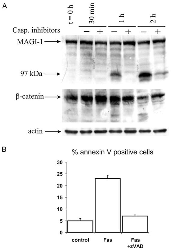
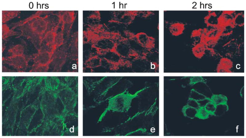
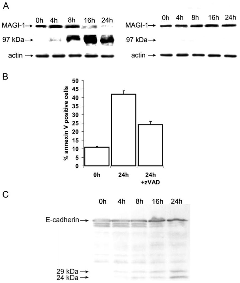
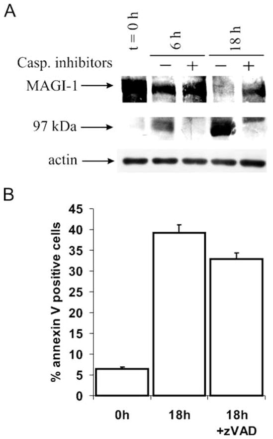
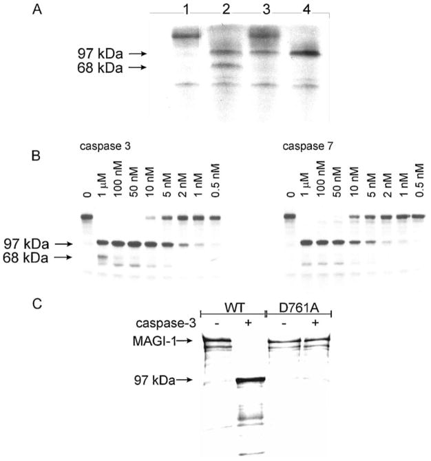
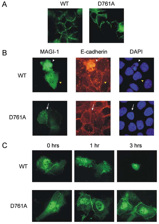
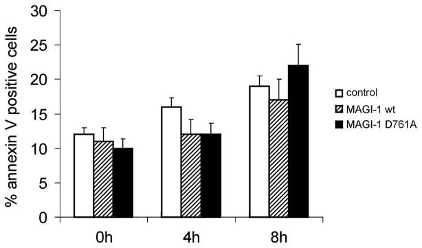
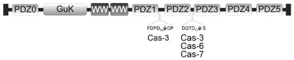
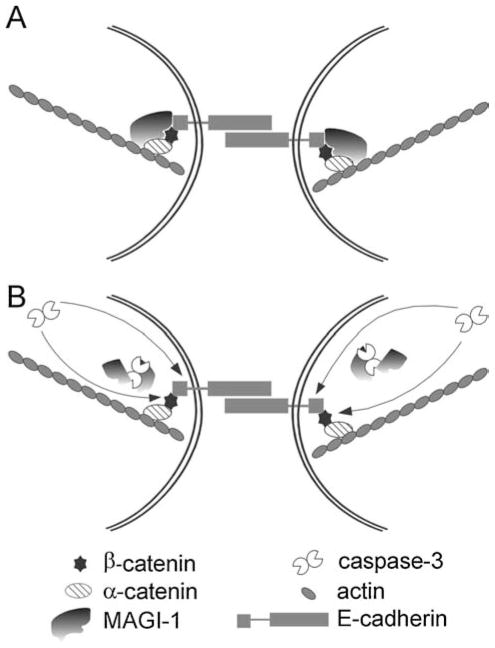
MAGI-1, a member of the MAGUK family of proteins, is shown to be rapidly cleaved during Fas-induced apoptosis in mouse 3T3 A31 cells, and in UV irradiation- and staurosporine-induced apoptosis in HaCaT cells. This generates a 97 kDa N-terminal fragment that dissociates from the cell membrane; a process that is largely prevented in the presence of the caspase inhibitor Z-VAD-fmk. In addition, we show that in vitro translated radiolabelled MAGI-1 is efficiently cleaved into 97 kDa and 68 kDa fragments by caspases-3 and -7 at physiological concentrations and mutating the MAGI-1 Asp(761) to Ala completely abolished the caspase-induced cleavage. Moreover, in HaCaT cells overexpressing the MAGI-1 Asp(761)Ala mutant the disruption of cell-cell contacts was delayed during apoptosis, whereas other caspase-dependent processes such as nuclear condensation were not affected, suggesting that cell detachment is parallel to them. Thus, MAGI-1 cleavage appears to be an important step in the disassembly of cell-cell contacts during apoptosis.
Figures
Similar articles
-
hDLG/SAP97, a member of the MAGUK protein family, is a novel caspase target during cell-cell detachment in apoptosis.Biol Chem. 2005 Jul;386(7):705-10. doi: 10.1515/BC.2005.082. Biol Chem. 2005. PMID: 16207092
-
Characterization of BAG3 cleavage during apoptosis of pancreatic cancer cells.J Cell Physiol. 2010 Jul;224(1):94-100. doi: 10.1002/jcp.22097. J Cell Physiol. 2010. PMID: 20232307
-
Endothelial adhesion molecule ESAM binds directly to the multidomain adaptor MAGI-1 and recruits it to cell contacts.Exp Cell Res. 2004 Oct 15;300(1):121-33. doi: 10.1016/j.yexcr.2004.07.010. Exp Cell Res. 2004. PMID: 15383320
-
Regulation and involvement in cancer and pathological conditions of MAGI1, a tight junction protein.Anticancer Res. 2014 Jul;34(7):3251-6. Anticancer Res. 2014. PMID: 24982328 Review.
-
A Potential Role for MAGI-1 in the Bi-Directional Relationship Between Major Depressive Disorder and Cardiovascular Disease.Curr Atheroscler Rep. 2024 Sep;26(9):463-483. doi: 10.1007/s11883-024-01223-5. Epub 2024 Jul 3. Curr Atheroscler Rep. 2024. PMID: 38958925 Free PMC article. Review.
Cited by
-
Age-associated genes in human mammary gland drive human breast cancer progression.Breast Cancer Res. 2020 Jun 15;22(1):64. doi: 10.1186/s13058-020-01299-2. Breast Cancer Res. 2020. PMID: 32539762 Free PMC article.
-
Upsetting the Balance: When Viruses Manipulate Cell Polarity Control.J Mol Biol. 2018 Sep 28;430(19):3481-3503. doi: 10.1016/j.jmb.2018.04.016. Epub 2018 Apr 20. J Mol Biol. 2018. PMID: 29680664 Free PMC article. Review.
-
MAGI1 Mediates eNOS Activation and NO Production in Endothelial Cells in Response to Fluid Shear Stress.Cells. 2019 Apr 27;8(5):388. doi: 10.3390/cells8050388. Cells. 2019. PMID: 31035633 Free PMC article.
-
MAGUKs, scaffolding proteins at cell junctions, are substrates of different proteases during apoptosis.Cell Death Dis. 2011 Jan 20;2(1):e116. doi: 10.1038/cddis.2010.92. Cell Death Dis. 2011. PMID: 21368887 Free PMC article.
-
Human papillomavirus E6 and E7: What remains?Tumour Virus Res. 2021 Jun;11:200213. doi: 10.1016/j.tvr.2021.200213. Epub 2021 Feb 8. Tumour Virus Res. 2021. PMID: 33716206 Free PMC article. Review.
References
-
- Steller H. Mechanism and genes of cellular suicide. Science. 1995;267:1445–1449. - PubMed
-
- Thompson CB. Apoptosis in the pathogenesis and treatment of disease. Science. 1995;267:1456–1462. - PubMed
-
- Nicholson DW, Thornberry NA. Caspases: killer proteases. Trends Biochem Sci. 1997;22:299–306. - PubMed
-
- Thornberry NA, Lazebnik Y. Caspases: enemies within. Science. 1998;281:1312–1316. - PubMed
-
- Salvesen GS, Dixit VM. Caspases: intracellular signaling by proteolysis. Cell. 1997;91:443–446. - PubMed
Publication types
MeSH terms
Substances
Grants and funding
LinkOut - more resources
Full Text Sources
Research Materials
Miscellaneous
