. 2007 Jan;57(1):90-102.
doi: 10.1002/mrm.21106.

Augmented generalized SENSE reconstruction to correct for rigid body motion

Affiliations

Augmented generalized SENSE reconstruction to correct for rigid body motion

Roland Bammer et al. Magn Reson Med. 2007 Jan.

Abstract

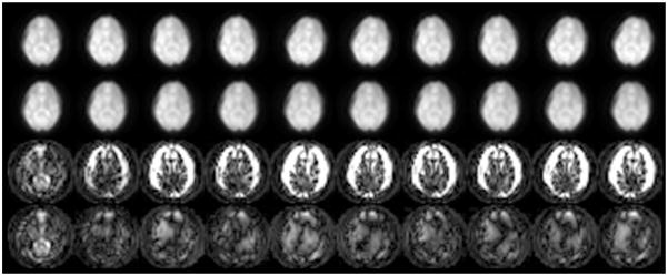
The correction of motion artifacts continues to be a significant problem in MRI. In the case of uncooperative patients, such as children, or patients who are unable to remain stationary, the accurate determination and correction of motion artifacts becomes a very important prerequisite for achieving good image quality. The application of conventional motion-correction strategies often produces inconsistencies in k-space data. As a result, significant residual artifacts can persist. In this work a formalism is introduced for parallel imaging in the presence of motion. The proposed method can improve overall image quality because it diminishes k-space inconsistencies by exploiting the complementary image encoding capacity of individual receiver coils. Specifically, an augmented version of an iterative SENSE reconstruction is used as a means of synthesizing the missing data in k-space. Motion is determined from low-resolution navigator images that are coregistered by an automatic registration routine. Navigator data can be derived from self-navigating k-space trajectories or in combination with other navigation schemes that estimate patient motion. This correction method is demonstrated by interleaved spiral images collected from volunteers. Conventional spiral scans and scans corrected with proposed techniques are shown, and the results illustrate the capacity of this new correction approach.


Figures

FIG. 1
FIG. 1
The effects of rotation are corrected by counter-rotating the individual interleaves. The effect of this counter-rotation on the k-space trajectories is shown for both interleaved spirals (a) and interleaved EPI (b). It is clearly apparent that the counter-rotation causes a variable sampling density in various areas of k-space. Some regions become oversampled compared to the Nyquist sampling rate, whereas other regions become only sparsely sampled. In the case of EPI, it is also worthwhile to note that the sampled trajectory data are no longer on an equidistant grid.
FIG. 2
FIG. 2
a: Axial GRE image acquired with one component coil (dashed line) from a dedicated head array coil. The coil is most sensitive to nearby regions and thus produces a highly nonuniform image. b: If the head is rotated in such a configuration, a different region of the object enters the most sensitive part of the coil, and hence the coil sensitivity has to be counter-rotated for correction purposes if the patient defines the frame of reference.
FIG. 3
FIG. 3
SE interleaved spiral pulse sequence and k-space trajectories used to obtain navigator images. a and b: A variable-density spiral design can be designed to cover enough information around the center of k-space so that a low-resolution navigator image can be produced. Alternatively, a short (~3–5 ms), single-shot, spiral-IN trajectory can be used during the initial SE formation followed by a conventional interleaved spiral trajectory to form the desired image.
FIG. 4
FIG. 4
Assessment of the effect of object rotation during an interleaved spiral data acquisition (eight interleaves) simulated in a quality phantom. Six receiver coils are distributed equally around the circumference of the phantom. For each interleaf, a random object rotation within the range of ±30° and a random object translation within the range of ±15 mm were introduced, with the following results: (a) gridding reconstruction of a quality phantom without rotation, (b) gridded k-space data, and (c) spiral sampling trajectory. Note that for better visualization of the sampling trajectory, only the region ±kmax/4 is plotted. If the acquired data are gridded according to the prescribed trajectory (c and f), inconsistencies in k-space (e) cause severe artifacts in the reconstructed image (d). Some of these distortions can be reduced (g) if the object rotation is used to counter-rotate the k-space acquisition trajectory for that particular interleaf (i), and these corrected orientations are used for gridding which corresponds to the operations on the left side of Eq. [8] (h). Correction for altered sensitivity and an iterative SENSE reconstruction (10 iterations) can remove most of the residual k-space sampling errors (k) and provides an image that is almost free of artifacts (j).
FIG. 5
FIG. 5
Assessment of the effect of object rotation during an interleaved with phase-encoding left-right EPI acquisition (eight interleaves with phase-encoding left-right) simulated in a quality phantom. Six receiver coils were distributed equally around the circumference of the phantom. For each interleaf, a random object rotation within the range of ±30° and a random object translation within the range of ±15 mm were introduced, with the following results: (a) gridding reconstruction of a quality phantom without rotation, (b) gridded k-space data, (c) EPI sampling trajectory, (d) gridding reconstruction of rotationally corrupted data, (e) corrupted k-space, (f) trajectory used for gridding, (g) gridding reconstruction of rotationally corrupted data using a rotationally corrected sampling trajectory, (h) gridded k-space with variable sampling density and regional undersampling, (i) rotationally corrected trajectory used for gridding, (j) resulting image after correction for altered coil sensitivity and iterative SENSE reconstruction (10 iterations), and (k) corresponding k-space.
FIG. 6
FIG. 6
In vivo experiment conducted with a fully sampled, low-resolution (32 × 32), single-shot, spiral-IN navigator preceding each conventional spiral interleaf. Shown are motion-corrected SE T2-weighted interleaved spiral-OUT scans with no motion (a), mild motion (b), and moderate motion (c). d–f: The resulting images reconstructed from the same data (i.e., no motion (d), mild motion (e), and moderate motion (f)) without any motion correction applied. e: Without correction, even mild motion causes significant artifacts that are mostly apparent in the frontal brain. This is because the origin of the rotation axis is located occipitally. f: Increased image artifacts can be seen with moderate motion. Without motion, there are no apparent differences between the reconstruction performed with (a) and without (d) motion correction. However, obvious artifact reduction can be achieved with the parallel imaging-based correction scheme in cases of mild (b vs. e) and moderate (c vs. f) motion. c: With moderate motion, some residual artifacts are apparent due to the severity of motion.
FIG. 7
FIG. 7
In vivo experiment conducted with a fully sampled, low-resolution (32 × 32), single-shot, spiral-IN navigator preceding each interleaf of a GRE-based interleaved spiral-OUT scan with TE = (a) 12 ms and (b) 90 ms. Top left: The reference sum-of-squares interleaved spiral image is acquired with no subject motion. Top right: Significant artifacts are apparent when the volunteer performs moderate head motion during data acquisition and no motion correction is applied. Bottom left: Correction of k-space data for translational and rotation motion results in sharper object contours, but the image quality is still corrupted by residual ghosting from local undersampling and sampling density variations. Bottom right: Image quality improves after the application of the augmented SENSE reconstruction, especially for the TE = 90 ms case (Fig. 7b). The SENSE-reconstructed image shows less artifacts than the conventionally reconstructed (i.e., gridding reconstruction) image with supposedly no motion.
FIG. 8
FIG. 8
Ten out of 32 navigator images obtained from consecutive acquisitions (32 interleaves). Top row: Navigator images before coregistration. Second row: Aligned navigator images after each image was registered to an average image computed from all 32 navigators. Third row: Absolute-squared-differences images (individual navigator images before registration vs. reference image). Spatial misregistration is clearly apparent from the hyperintense rim at the edge of the brain. The intensity scaling is increased 10-fold compared to the two rows to better highlight the differences. Bottom row: Absolute-squared-differences images (individual navigator images after registration vs. reference image) clearly demonstrate a better alignment. The scaling is identical to that of the images in the third row.

References

    1. Larkman DJ, Atkinson D, Hajnal JV. Artifact reduction using parallel imaging methods. Top Magn Reson Imaging. 2004;15:267–275. - PubMed
    1. Pipe JG, Farthing VG, Forbes KP. Multishot diffusion-weighted FSE using PROPELLER MRI. Magn Reson Med. 2002;47:42–52. - PubMed
    1. Bydder M, Larkman DJ, Hajnal JV. Detection and elimination of motion artifacts by regeneration of k-space. Magn Reson Med. 2002;47:677– 686. - PubMed
    1. Atkinson D, Hill DL. Reconstruction after rotational motion. Magn Reson Med. 2003;49:183–187. - PubMed
    1. Bydder M, Atkinson D, Larkman DJ, Hill DL, Hajnal JV. SMASH navigators. Magn Reson Med. 2003;49:493–500. - PubMed

Publication types