. 2007 Mar;81(6):2805-16.
doi: 10.1128/JVI.02095-06. Epub 2006 Dec 27.

Complete genome analysis of 33 ecologically and biologically diverse Rift Valley fever virus strains reveals widespread virus movement and low genetic diversity due to recent common ancestry

Affiliations

Complete genome analysis of 33 ecologically and biologically diverse Rift Valley fever virus strains reveals widespread virus movement and low genetic diversity due to recent common ancestry

Brian H Bird et al. J Virol. 2007 Mar.

Abstract

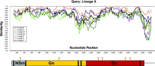
Rift Valley fever (RVF) virus is a mosquito-borne RNA virus responsible for large explosive outbreaks of acute febrile disease in humans and livestock in Africa with significant mortality and economic impact. The successful high-throughput generation of the complete genome sequence was achieved for 33 diverse RVF virus strains collected from throughout Africa and Saudi Arabia from 1944 to 2000, including strains differing in pathogenicity in disease models. While several distinct virus genetic lineages were determined, which approximately correlate with geographic origin, multiple exceptions indicative of long-distance virus movement have been found. Virus strains isolated within an epidemic (e.g., Mauritania, 1987, or Egypt, 1977 to 1978) exhibit little diversity, while those in enzootic settings (e.g., 1970s Zimbabwe) can be highly diverse. In addition, the large Saudi Arabian RVF outbreak in 2000 appears to have involved virus introduction from East Africa, based on the close ancestral relationship of a 1998 East African virus. Virus genetic diversity was low (approximately 5%) and primarily involved accumulation of mutations at an average of 2.9 x 10(-4) substitutions/site/year, although some evidence of RNA segment reassortment was found. Bayesian analysis of current RVF virus genetic diversity places the most recent common ancestor of these viruses in the late 1800s, the colonial period in Africa, a time of dramatic changes in agricultural practices and introduction of nonindigenous livestock breeds. In addition to insights into the evolution and ecology of RVF virus, these genomic data also provide a foundation for the design of molecular detection assays and prototype vaccines useful in combating this important disease.


Figures

FIG. 1.
FIG. 1.
Simplot analysis of 33 RVF virus complete M segment nucleotide alignments grouped by virus lineage (A to G), indicating nucleotide positions (antigenomic sense) along the x axis and, on the y axis, percent nucleotide similarity. Depicted beneath the x axis is a cartoon representation of the 5′ UTR; the NSm, Gn, and Gc coding regions; and the 3′ UTR encoded by the RVF virus M segment. Potential glycosylation sites are indicated above by arrows. Predicted hydrophobic transmembrane domains are depicted by heavy black lines. Simplot analyses (sliding window, 180 nt; step, 20 nt) revealed regions of greater nucleotide diversity, approaching 20% at the carboxy terminus of Gn and the amino terminus of Gc and at the 3′ UTR.
FIG. 2.
FIG. 2.
Thirty-three complete RVF S segments were analyzed by either unconstrained ML techniques with 500 replicate bootstrap values (PAUP*b10) or the Bayesian statistical program (BEAST) with an MCMC chain length of 3.0 × 10⁷, a 3.0 × 10⁶ burn in, a GTR+Γ+I nucleotide rate substitution model, a strict molecular clock, and sampling of every 1,000 states. A strict molecular clock was chosen after multiple analyses; using both relaxed exponential and relaxed logarithmic clock models revealed no significant deviation in the coalescence of the overall evolutionary rate or tree topologies. Trees and support values from the ML and BEAST analyses were similar. The Bayesian MAP log likelihood value tree was chosen from the posterior distribution and is depicted here. Posterior support values were calculated from consensus analysis of all Bayesian posterior trees, with values over 0.5 indicated above each node. The estimated TMRCA of the tree nodes was calculated by Bayesian analyses and is reported below each node as years prior to the year 2000. The overall TMRCA of the entire S segment analysis is indicated in a text box located adjacent to the root node of SA51. Each taxon name indicates the strain, country of origin, and date of isolation. The GenBank accession numbers for the virus S segments are DQ380143 to -6, DQ380149, DQ380151 to -3, DQ380156, and DQ380158 to -81. Strains used in previous studies of virulence in WF rats are indicated with either + (lethal; LD50, ∼1.0 PFU), +/−, (lethal; LD50, ∼2 × 10³ PFU), or − (nonlethal).
FIG. 3.
FIG. 3.
Thirty-three complete RVF M segments were analyzed by either unconstrained ML techniques with 500 replicate bootstrap values (PAUP*b10) or the Bayesian statistical program (BEAST) with an MCMC chain length of 3.0 × 10⁷, a 3.0 × 10⁶ burn in, a GTR+Γ+I nucleotide rate substitution model, a strict molecular clock, and sampling of every 1,000 states. Trees and support values from the ML and BEAST analyses were similar. The Bayesian MAP log likelihood value tree was chosen from the posterior distribution and is depicted here. Posterior support values were calculated from consensus analysis of all Bayesian posterior trees, with values over 0.5 indicated above each node. The estimated TMRCA of tree nodes was calculated by Bayesian analyses and is reported below each node as years prior to the year 2000. The overall TMRCA of the entire M segment analysis is indicated in a text box located adjacent to the root node of SA51. Each taxon name indicates the strain, country of origin, and date of isolation. GenBank accession numbers for the virus M segments are DQ380183 to -91, DQ380194 to -8, DQ380200, DQ380203 to -7, DQ380209 to -12, and DQ380214 to -22. Strains used in previous studies of virulence in WF rats are indicated with either + (lethal; LD50, ∼1.0 PFU), +/− (lethal; LD50, ∼2 × 10³ PFU), or − (nonlethal).
FIG. 4.
FIG. 4.
Thirty-three complete RVF L segments were analyzed by either unconstrained ML techniques with 500 replicate bootstrap values (PAUP*b10) or the Bayesian statistical program (BEAST) with an MCMC chain length of 3.0 × 10⁷, a 3.0 × 10⁶ burn in, a GTR+Γ+I nucleotide rate substitution model, a strict molecular clock, and sampling of every 1,000 states. Trees and support values from the ML and BEAST analyses were similar. The Bayesian MAP log likelihood value tree was chosen from the posterior distribution and is depicted here. Posterior support values were calculated from consensus analysis of all Bayesian posterior trees, with values over 0.5 indicated above each node. The estimated TMRCA of tree nodes was calculated by Bayesian analyses and is reported below each node as years prior to the year 2000. The overall TMRCA of the entire L segment analysis is indicated in a text box located adjacent to the root node of SA51. Each taxon name indicates the strain, country of origin, and date of isolation. GenBank accession numbers for the virus L segments are DQ375395 to -403, DQ375406, DQ375409 to -16, DQ375418 to -29, and DQ375432 to -4. Strains used in previous studies of virulence in WF rats are indicated with either + (lethal; LD50, ∼1.0 PFU), +/− (lethal; LD50, ∼2 × 10³ PFU), or − (nonlethal).

Similar articles

Cited by

References

    1. Anderson, G. W., Jr., W. Lawrence, J. O. Lee, and William C. Hall. Ocular sequelae associated with Rift Valley fever virus (RVFV) infection in inbred rat strains. Abstr. VIIIth Int. Congr. Virol., abstr. P70-002.
    1. Anderson, G. W., Jr., and C. J. Peters. 1988. Viral determinants of virulence for Rift Valley fever (RVF) in rats. Microb. Pathog. 5:241-250. - PubMed
    1. Anderson, G. W., Jr., J.-F. Saluzzo, T. G. Ksiazek, J. F. Smith, W. Ennis, D. Thureen, C. J. Peters, and J.-P. Digoutte. 1989. Comparison of in vitro and in vivo systems for propagation of Rift Valley fever virus from clinical specimens. Res. Virol. 140:129-138. - PubMed
    1. Aquino, V. H., M. L. Moreli, and L. T. Moraes Figueiredo. 2003. Analysis of oropouche virus L protein amino acid sequence showed the presence of an additional conserved region that could harbour an important role for the polymerase activity. Arch. Virol. 148:19-28. - PubMed
    1. Beaty, B. J., D. R. Sundin, L. J. Chandler, and D. H. Bishop. 1985. Evolution of bunyaviruses by genome reassortment in dually infected mosquitoes (Aedes triseriatus). Science 230:548-550. - PubMed

Publication types

MeSH terms

LinkOut - more resources