. 2007 Mar;81(6):2899-908.
doi: 10.1128/JVI.01850-06. Epub 2006 Dec 27.

Nuclear import of bovine papillomavirus type 1 E1 protein is mediated by multiple alpha importins and is negatively regulated by phosphorylation near a nuclear localization signal

Affiliations

Nuclear import of bovine papillomavirus type 1 E1 protein is mediated by multiple alpha importins and is negatively regulated by phosphorylation near a nuclear localization signal

Xue-Lin Bian et al. J Virol. 2007 Mar.

Abstract

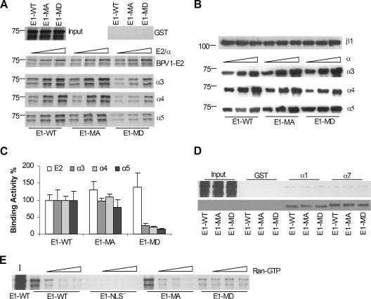
Papillomavirus DNA replication occurs in the nucleus of infected cells and requires the viral E1 protein, which enters the nuclei of host epithelial cells and carries out enzymatic functions required for the initiation of viral DNA replication. In this study, we investigated the pathway and regulation of the nuclear import of the E1 protein from bovine papillomavirus type 1 (BPV1). Using an in vitro binding assay, we determined that the E1 protein interacted with importins alpha3, alpha4, and alpha5 via its nuclear localization signal (NLS) sequence. In agreement with this result, purified E1 protein was effectively imported into the nucleus of digitonin-permeabilized HeLa cells after incubation with importin alpha3, alpha4, or alpha5 and other necessary import factors. We also observed that in vitro binding of E1 protein to all three alpha importins was significantly decreased by the introduction of pseudophosphorylation mutations in the NLS region. Consistent with the binding defect, pseudophosphorylated E1 protein failed to enter the nucleus of digitonin-permeabilized HeLa cells in vitro. Likewise, the pseudophosphorylation mutant showed aberrant intracellular localization in vivo and accumulated primarily on the nuclear envelope in transfected HeLa cells, while the corresponding alanine replacement mutant displayed the same cellular location pattern as wild-type E1 protein. Collectively, our data demonstrate that BPV1 E1 protein can be transported into the nucleus by more than one importin alpha and suggest that E1 phosphorylation by host cell kinases plays a regulatory role in modulating E1 nucleocytoplasmic localization. This phosphoregulation of nuclear E1 protein uptake may contribute to the coordination of viral replication with keratinocyte proliferation and differentiation.


Figures

FIG. 1.
FIG. 1.
Papillomavirus E1 proteins bind α importins in vitro. (A) The sequence of WT BPV1 E1 protein in the region of the NLS is shown, with the clusters of basic residues underlined. The amino acid changes present in three mutant E1 proteins (NLS; a double pseudophosphorylation mutant, MD; and the uncharged double-alanine mutant MA) constructed for this study are indicated below the WT E1 sequence. (B) Autoradiograph of the in vitro-translated HA-E1-WT and HA-E1-NLS proteins bound to His-Com1 (2.0 μg) or increasing amounts (0.5, 1.0, and 2.0 μg) of different importin α proteins (indicated by the hollow triangles). The input panel shows a sample of the in vitro-translated E1 proteins used for the binding assay. (C) Immunoblot of the His-importin α fusion proteins used in the pull-down assay shown in B. His-importins were detected with an anti-His antibody. (D) Quantification of the full-length WT E1 product (75 kDa) bound to His-Com1 and the different importin α proteins by phosphordensitometry. The amount of bound NLS E1 was subtracted from the amount of bound WT E1 for Com1 and each of the five α importins tested, and the difference is presented as the percentage of the input WT E1 protein. Error bars represent standard errors obtained in two or three experiments. (E) Autoradiograph of the in vitro-translated HPV11 E1 bound to His-Com1 (2.0 μg) or increasing amounts (0.5, 1.0, and 2.0 μg) of different importin α proteins (upper panel). The assay was performed as described above for BPV1 E1. The lower panel shows the immunoblot of the importins as in C. Numbers to the left of the blots and autoradiographs in B, C, and E show the positions of molecular mass markers (in kilodaltons). (F) Immunoblot of GST-mCry2 bound to 1.0 μg of importin α7. Equal amounts of GST or GST-mCry2 were incubated with α7, the complexes were collected on Ni-NTA agarose, and the bound material was detected with anti-GST. (G) Coimmunoprecipitation of E1 with α importin. Extracts from cells expressing either eGFP or eGFP-E1 were immunoprecipitated with anti-importin α4 and then immunoblotted with anti-GFP or anti-importin α4 as indicated.
FIG. 2.
FIG. 2.
The BPV1 E1 protein is imported into the nuclei of permeabilized cells by importins α3, α4, and α5 but not by importins α1 and α7. (A) Coomassie blue staining of 0.5 μg of the purified GST-NLS, GST-E11-311-WT, and GST-E11-311-NLS proteins used for the in vitro uptake assay. The positions of molecular mass markers (in kilodaltons) are indicated on the left side of each panel. (B) Nuclear import assay of the fluorescence-labeled GST-NLS fusion protein (positive control) tested in the absence or presence of different importin α proteins as indicated. The assay was performed as described in Materials and Methods, and the labeled GST-NLS protein was detected by fluorescence microscopy. (C) Nuclear import assay as in B with fluorescence-labeled GST-E11-311-WT and GST-E11-311-NLS.
FIG. 3.
FIG. 3.
Pseudophosphorylation of the BPV1 E1 protein at T102 and S109 reduces its binding activity to importin α proteins in vitro. (A) Autoradiograph of the in vitro-translated His-E1-WT, His-E1-MA, and His-E1-MD proteins (expressed from pRSET) bound to α importins in the presence of GST (upper right panel) or GST-importin β1 (α3, α4, and α5 panels) or to GST-E2 alone (BPV1-E2 panel) as described in Materials and Methods. Hollow triangles indicate increasing amounts of α importins or BPV1 E2 used in the binding reaction mixtures (0.5, 1.0, and 2.0 μg). The input panel (upper left) shows a sample of the in vitro-translated WT and mutant E1 proteins. The position of a 75-kDa molecular mass marker is indicated to the left. (B) Immunoblots of importins α and β1 used in pull-down assay shown in A. Proteins were detected with anti-His (α importins) or anti-GST (β importin) antibody. (C) Phosphordensitometric quantification of the full-length E1 product (75 kDa) bound to α importins in the presence of importin β1 or bound to BPV1 E2 protein. Data shown in the graph are derived from binding reactions shown in A using 2.0 μg of wild-type or mutant E1 proteins. The binding activities of the E1-MA and E1-MD mutants are relative to that of the WT BPV1 E1 protein, which was assigned as 100%. Error bars represent standard errors obtained in two or three experiments. (D) The upper panel is an autoradiograph of in vitro-translated WT or mutant E1 protein bound to complexes of α1/GST-β1, α7/GST-β1, or GST alone. The input lanes show portions of the original in vitro translation reaction for the WT, MA, and MD E1 proteins as indicated. The lower panel is an immunoblot of α importins used in the pull-down assay shown in the upper panel. The importins were detected with anti-His antibody. (E) The E1 binding assay was performed with WT and mutant E1 proteins as in A except with the inclusion of 0.5, 1.0, or 2.0 μg of Ran-GTP in the reaction mixture, as indicated by the hollow triangles. The lane marked I shows the input WT E1 protein.
FIG. 4.
FIG. 4.
Pseudophosphorylation of BPV1 E1 at T102 and S109 affects BPV1 E1 protein nuclear import. (A) Nuclear uptake of GST-E11-311-WT, GST-E11-311-MA, and GST-E11-311-MD in permeabilized HeLa cells supplemented with importins α3, α4, and α5 as indicated. The assay was performed as described in the legend of Fig. 2, and the localization of the E1 protein was detected by fluorescence microscopy. (B) Coomassie blue staining of 0.5 μg of GST-E11-311-WT, GST-E11-311-MA, and GST-E11-311-MD proteins used in the in vitro import assay, with the molecular mass markers (in kilodaltons) indicated on the left. (C) HeLa cells were transfected with peGFP-E1-WT, peGFP-E1-MA, peGFP-E1-MD, and pREV1-4(NES3)GFP (positive control). The cellular localization of the E1 protein was detected 24 h after transfection by fluorescence microscopy for the eGFP signal.
FIG. 5.
FIG. 5.
Examination of other possible posttranslational modification sites of BPV1 E1 for involvement in E1 binding to importin α. Wild-type E1 and the four pseudophosphorylation mutants were all expressed in vitro from pRSET. (A) Autoradiograph of the in vitro-translated WT and mutant E1 proteins bound to importin β1/α5 complexes or to GST alone as described in the legend of Fig. 3. Hollow triangles indicate increasing amounts of importin α5 (0.5, 1.0, and 2.0 μg) used in the binding reaction mixtures (for GST, only the results with 2 μg are shown). The input lanes show samples of the original in vitro-translated WT and mutant E1 proteins used for the binding reactions. Two independent experiments (upper and lower panels) are shown, with the position of the 75-kDa molecular mass marker indicated on the left. (B) Phosphordensitometry quantification of the bound WT and mutant E1 proteins calculated as described in the legend of Fig. 3C. (C) Tabular presentation of the quantitative data shown graphically in B.

References

    1. Adam, S. A., R. S. Marr, and L. Gerace. 1990. Nuclear protein import in permeabilized mammalian cells requires soluble cytoplasmic factors. J. Cell Biol. 111:807-816. - PMC - PubMed
    1. Alvisi, G., D. A. Jans, J. Guo, L. A. Pinna, and A. Ripalti. 2005. A protein kinase CK2 site flanking the nuclear targeting signal enhances nuclear transport of human cytomegalovirus ppUL44. Traffic 6:1002-1013. - PubMed
    1. Angeline, M., E. Merle, and J. Moroianu. 2003. The E7 oncoprotein of high-risk human papillomavirus type 16 enters the nucleus via a nonclassical Ran-dependent pathway. Virology 317:13-23. - PubMed
    1. Blachon, S., S. Bellanger, C. Demeret, and F. Thierry. 2005. Nucleo-cytoplasmic shuttling of high risk human papillomavirus E2 proteins induces apoptosis. J. Biol. Chem. 280:36088-36098. - PubMed
    1. Bodily, J. M., S. Alam, and C. Meyers. 2006. Regulation of human papillomavirus type 31 late promoter activation and genome amplification by protein kinase C. Virology 348:328-340. - PubMed

Publication types

MeSH terms

LinkOut - more resources