. 2007 Mar;21(3):613-24.
doi: 10.1210/me.2006-0269. Epub 2006 Dec 27.

Proline-, glutamic acid-, and leucine-rich protein-1/modulator of nongenomic activity of estrogen receptor enhances androgen receptor functions through LIM-only coactivator, four-and-a-half LIM-only protein 2

Affiliations

Proline-, glutamic acid-, and leucine-rich protein-1/modulator of nongenomic activity of estrogen receptor enhances androgen receptor functions through LIM-only coactivator, four-and-a-half LIM-only protein 2

Sujit S Nair et al. Mol Endocrinol. 2007 Mar.

Abstract

Proline-, glutamic acid-, and leucine-rich protein-1 (PELP1) is a coregulator of multiple nuclear receptors. Molecular mechanisms of PELP1 function are not completely understood, but its expression is up-regulated in hormonal-dependent cancers. Using a yeast two-hybrid screen, we found that four-and-a-half LIM-only protein 2 (FHL2) interacted with PELP1. FHL2 is a transcriptional regulator that associates with nuclear cofactors, including androgen receptors (ARs), and contains an intrinsic activation domain. PELP1 and FHL2 interact in vitro and in vivo and colocalize in the nuclear compartment. PELP1 interacts with FHL2 via LIM domains 3 and 4 and synergistically enhances the transcriptional activity of FHL2. Src kinase is required for PELP1-mediated enhancement of FHL2 functions because knockdown of Src kinase expression or function abolished PELP1-mediated FHL2 activation functions. PELP1 interacted with AR and enhanced FHL2-mediated AR transactivation functions. PELP1 knockdown by small interfering RNA or PELP1 mutant, which lacks an activation domain, reduced FHL2-mediated AR transactivation. Biochemical analyses revealed a complex consisting of PELP1, FHL2, and AR in prostate cancer cells. PELP1/MNAR expression was elevated in high-grade prostate tumors. Our results suggest that PELP1 functions as a molecular adaptor, coupling FHL2 with nuclear receptors, and PELP1-FHL2 interactions may have a role in prostate cancer progression.


Figures

Fig. 1
Fig. 1. PELP1/MNAR Interacts with FHL2
A, Yeast cells were transfected with a control Gal4-activation domain (GAD) vector or GAD-FHL2, along with a Gal4-DNA binding domain (GBD) vector or GBD-PELP1/MNAR. Growth was recorded after 72 h on selection plates lacking leucine and tryptophan (−LT) or adenosine, histidine, leucine, and tryptophan (−AHLT). B, HEK293 cells were transfected with T7-PELP1/MNAR and Flag-FHL2. Immunoprecipitation was performed with a Flag antibody, followed by Western blot analysis with a T7 antibody. C, Nuclear extracts from HeLa cells were subjected to immunoprecipitation using a control IgG or a PELP1/MNAR antibody, followed by Western blot analysis using an FHL2 antibody. D, HeLa cells grown in 10% serum were fixed and costained with antibodies against PELP1/MNAR (green) and FHL2 (red). DNA is stained blue by Topro-3 (TOPRO). The images were analyzed by confocal microscopy. Colocalization of PELP1/MNAR and FHL2 is shown in yellow. IP, Immunoprecipitation.
Fig. 2
Fig. 2. PELP1/MNAR Is a Coactivator of FHL2-Dependent Transcription
A, Total cell lysates from COS-7 cells expressing T7-PELP1/MNAR were incubated with GST-FHL2 fusion proteins of various lengths, and the PELP1/MNAR binding region in FHL2 was analyzed with a GST pull-down assay. B, PELP1 peptides of indicated lengths were translated in vitro using the TNT system in the presence of [35S]methionine, and their ability to bind GST-FHL2 was analyzed by GST pull-down analysis. C, PC3 cells were cotransfected with a Gal4-based Luc reporter (G5E1b-Luc), β-Gal reporter, Gal4-FHL2 with or without PELP1/MNAR. After 48 h, G5E1b-Luc reporter activity was measured. D, PC3 cells were cotransfected with G5E1b-Luc, β-Gal reporter and Gal4FHL2 with or without full-length or with serial deletions of PELP1/MNAR. After 48 h, G5E1b-Luc reporter activity was measured. In all the reporter gene assays parental vectors were used as controls and the total amount of the DNA in the transfections was kept constant by adding appropriate vectors. β-Gal values were used to normalize luciferase activity for transfection efficiency. Data shown are the means of ± SE from three independent experiments performed in triplicate wells. *, P < 0.05.
Fig. 3
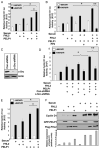
Fig. 3. PELP1 Modulates FHL2-Mediated Target Gene Activation in an Src-Dependent Manner
HeLa cells were cotransfected with a cyclin D1-Luc reporter with or without PELP1/MNAR and with or without FHL2. A, After serum starvation for 48 h, cells were stimulated with serum (10%) for 12 h, and luciferase activity was measured. B, After 48 h, cells were pretreated with an Src inhibitor PP2 (10 μM, for 1 h) and then stimulated with serum for 12 h. C, Down-regulation of c-Src expression by shRNA was analyzed by Western analysis. D, At 24 h after transfection, indicated wells were infected with control or c-Src-shRNA-specific lentivirus. After 72 h of virus infection, cells were treated with or without serum, and luciferase activity was measured. In all the reporter gene assays parental vectors were used as controls, and the total amount of the DNA in the transfection was kept constant by adding appropriate empty vectors. β-Gal values were used to normalize luciferase activity for transfection efficiency. Data shown are the means ± SE from three independent experiments performed in triplicate wells. *, P < 0.05 comparing PELP1+FHL2 activity vs. PELP1 or FHL2. **, P < 0.001 comparing PELP1 vs. PP2 or c-Src-shRNA. E, NIH 3T3 cells were cotransfected with a cyclin D1-Luc reporter, β-Gal reporter gene, with or without PELP1/MNAR and with or without FHL2. After serum starvation for 48 h, cells were stimulated with serum (10%) for 12 h, and luciferase activity was measured. F, NIH 3T3 cells were transfected with GFP or GFP-PELP1 vector along with or without flag-FHL2 vector. After 48 h cells were subjected to fluorescence-activated cell sorting for GFP expression. Equal number of GFP or GFP PELP1 sorted cells were cultured in duplicate plates. After 48 h, cells were treated with or without serum for 12 h and cyclin D1 levels were analyzed by Western blotting. Con-shRNA, Control shRNA.
Fig. 4
Fig. 4. PELP1/MNAR Enhances FHL2-Mediated AR Transactivation
A, PC3 cells were cotransfected with MMTV-Luc, β-Gal reporter gene, and AR, with or without PELP1/MNAR and with or without FHL2. After 48 h, cells were treated with R1881, and MMTV-Luc reporter activity was measured. B, PC3 cells were cotransfected with MMTV-Luc, β-Gal reporter gene, with or without PELP1/MNAR and with or without FHL2, and after 48 h, reporter gene activation was measured. C, PC3 cells were cotransfected with MMTV-Luc, β-Gal reporter gene, AR, and FHL2 with PELP1/MNAR-1–600 or PELP1/MNAR-800–1130. After 48 h, cells were treated with R1881 for 12 h, and luciferase activity was measured. D, PC3 cells were transfected with control or PELP1/MNAR-specific siRNA. After 24 h, they were transfected with MMTV-Luc, AR, and FHL2. After 48 h, cells were treated with R1881 for 12 h, and luciferase activity was measured. E, PC3 cells were cotransfected with MMTV-Luc, β-Gal reporter gene, and AR, with or without FHL2. After 48 h, cells were pretreated with the c-Src inhibitor, PP2 (10 μM for 1 h), before ligand treatment. After 48 h, cells were treated with R1881 for 12 h, and luciferase activity was measured. β-Gal values were used to normalize luciferase activity for transfection efficiency. Data shown are the means ± SE from three independent experiments performed in triplicate wells. *, P < 0.05 comparing PELP1+FHL2 activity vs. PELP1 or FHL2. **, P < 0.001 comparing FHL2 activation in the presence or absence of PELP1/MNAR siRNA. GAPDH, Glyceraldehyde-3-phosphate dehydrogenase.
Fig. 5
Fig. 5. PELP1/MNAR, AR, and FHL2 Form Functional Complexes in Vivo
A, LNCaP cells were cotransfected with a PSA-Luc reporter, with or without PELP1/MNAR and with or without FHL2. After 48 h, cells were treated with R1881 for 12 h, and luciferase activity was measured. *, P < 0.05. B, PC3(AR)2 cells were biochemically fractionated into nuclear and cytoplasmic extracts, and the total lysates were analyzed for PELP1/MNAR by Western analysis. Lamin B1 and paxillin were used as markers of the nucleus and cytoplasm, respectively. C, AR was immunoprecipitated from PC3(AR)2 cytoplasmic and nuclear lysates, and the presence of PELP1/MNAR in the immunoprecipitates was analyzed by Western blotting. D, LNCaP cells were treated with R1881, and nuclear extracts were prepared and subjected to immunoprecipitation with control IgG or AR antibody. The presence of PELP1/MNAR in the immunoprecipitates was analyzed by Western analysis. E, HEK293 cells coexpressing T7-PELP1/MNAR, Flag-FHL2, and AR were treated with 1% formaldehyde to stabilize the complexes, and cellular lysates were immunoprecipitated with T7 antibody. The presence of AR and FHL2 in the immunoprecipitates was analyzed by Western analysis. F, HEK293 cells were transfected with AR and Flag-FHL2 and with or without PELP1/MNAR. After 72 h, cells were treated with R1881 for 1 h, and the cell lysates were immunoprecipitated with a Flag antibody. The presence of AR in the immunoprecipitates was analyzed by Western blotting. G, HEK293 cells were transfected with AR and Flag-FHL2 with increasing amounts of PELP1/MNAR. After 72 h, cells were treated with R1881 for 1 h, and the cell lysates were immunoprecipitated with a Flag antibody. The presence of AR in the immunoprecipitates was analyzed by Western blotting. IP, Immunoprecipitation.
Fig. 6
Fig. 6. Expression of PELP1/MNAR in Prostate Cancer
Immunohistological analysis of PELP1/MNAR in a prostate tumor using an AccuMax tissue array and an affinity-purified PELP1 antibody (Bethyl Laboratories). A, Nonneoplastic; B, Gleason grade 5; C, Gleason grade 8; D, quantitation of the PELP1/MNAR expression in benign, low-grade (Gleason 4–6) and high-grade tumors (Gleason 7–9). Scoring was done as follows: weak/focal (1+), moderate (+2), and strong (+3). *, P < 0.05. Non, Nonneoplastic.

Similar articles

Cited by

References

    1. Vadlamudi RK, Wang RA, Mazumdar A, Kim Y, Shin J, Sahin A, Kumar R. Molecular cloning and characterization of PELP1, a novel human coregulator of estrogen receptor α. J Biol Chem. 2001;276:38272–38279. - PubMed
    1. Wong CW, McNally C, Nickbarg E, Komm BS, Cheskis BJ. Estrogen receptor-interacting protein that modulates its nongenomic activity-crosstalk with Src/Erk phosphorylation cascade. Proc Natl Acad Sci USA. 2002;99:14783–14788. - PMC - PubMed
    1. Nair SS, Mishra SK, Yang Z, Balasenthil S, Kumar R, Vadlamudi RK. Potential role of a novel transcriptional coactivator PELP1 in histone H1 displacement in cancer cells. Cancer Res. 2004;64:6416–6423. - PubMed
    1. Vadlamudi RK, Manavathi B, Balasenthil S, Nair SS, Yang Z, Sahin AA, Kumar R. Functional implications of altered subcellular localization of PELP1 in breast cancer cells. Cancer Res. 2005;65:7724–7732. - PMC - PubMed
    1. Vadlamudi RK, Balasenthil S, Broaddus RR, Gustafsson JA, Kumar R. Deregulation of estrogen receptor coactivator proline-, glutamic acid-, and leucine-rich protein-1/modulator of nongenomic activity of estrogen receptor in human endometrial tumors. J Clin Endocrinol Metab. 2004;89:6130–6138. - PMC - PubMed

Publication types

MeSH terms