. 2007 Mar;18(3):839-49.
doi: 10.1091/mbc.e06-08-0707. Epub 2006 Dec 27.

Two mammalian Sec16 homologues have nonredundant functions in endoplasmic reticulum (ER) export and transitional ER organization

Affiliations

Two mammalian Sec16 homologues have nonredundant functions in endoplasmic reticulum (ER) export and transitional ER organization

Dibyendu Bhattacharyya et al. Mol Biol Cell. 2007 Mar.

Abstract

Budding yeast Sec16 is a large peripheral endoplasmic reticulum (ER) membrane protein that functions in generating COPII transport vesicles and in clustering COPII components at transitional ER (tER) sites. Sec16 interacts with multiple COPII components. Although the COPII assembly pathway is evolutionarily conserved, Sec16 homologues have not been described in higher eukaryotes. Here, we show that mammalian cells contain two distinct Sec16 homologues: a large protein that we term Sec16L and a smaller protein that we term Sec16S. These proteins localize to tER sites, and an N-terminal region of each protein is necessary and sufficient for tER localization. The Sec16L and Sec16S genes are both expressed in every tissue examined, and both proteins are required in HeLa cells for ER export and for normal tER organization. Sec16L resembles yeast Sec16 in having a C-terminal conserved domain that interacts with the COPII coat protein Sec23, but Sec16S lacks such a C-terminal conserved domain. Immunoprecipitation data indicate that Sec16L and Sec16S are each present at multiple copies in a heteromeric complex. We infer that mammalian cells have preserved and extended the function of Sec16.


Figures
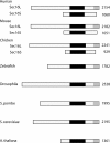
Figure 1.
Figure 1.
Schematic representations of Sec16 homologues from different species. Diagrammed are putative homologues of Sec16 from human (H. sapiens), mouse (M. musculus), chicken (G. gallus), zebrafish (D. rerio), fruit fly (D. melanogaster), fission yeast (S. pombe), budding yeast (S. cerevisiae), and mustard plant (A. thaliana). The black box represents the central conserved domain, and the gray box represents the C-terminal conserved domain. Listed on the right are the sizes of the predicted protein products in amino acids.
Figure 2.
Figure 2.
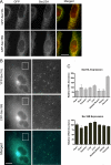
Sec16L and Sec16S colocalize with a COPII coat protein and with each other and are both expressed in a variety of tissues. (A) Colocalization of the Sec16 homologues with Sec23A. A plasmid encoding GFP-Sec16L (top) or GFP-Sec16S (bottom) was transiently transfected into HeLa cells. At 12 h after transfection, the cells were processed for immunofluorescence by using anti-GFP mAb plus anti-Sec23A polyclonal antibody. The merged images show colocalization of GFP (green) with Sec23A (red). Bar, 20 μm. (B) Colocalization of the Sec16 homologues with each other. A plasmid encoding YFP-Sec16L was cotransfected into HeLa cells with a plasmid encoding CFP-Sec16S. At 12 h after transfection, the cells were fixed and subjected to fluorescence microscopy. The panels on the right show an enlarged view of the inset in the panels on the left. The merged images show colocalization of YFP-Sec16L (green) with CFP-Sec16S (blue). Bar, 10 |gmm. (C) Tissue mRNA levels of the Sec16 homologues. Total RNA samples from the indicated human tissues were subjected to real-time RT-PCR by using primers specific for Sec16L or Sec16S. The linear phase yields were normalized against actin mRNA signals. The resulting numbers were then normalized again by defining the average signal from all of the tissues as 100. Bars indicate SEs of the mean calculated from three separate experiments.
Figure 3.
Figure 3.
RNAi-mediated knockdown of Sec16L or Sec16S disrupts tER sites. (A) Quantitation of RNAi-mediated knockdowns. HeLa cells cultured on coverslips were transfected either with a control nonspecific RNAi or with an RNAi against Sec16L, Sec16S, or Sec12. At 36 h after transfection, total RNA from each sample was subjected to real-time RT-PCR by using primers specific for Sec16L, Sec16S, or Sec12. The linear phase yields were normalized against actin mRNA signals, and the signals obtained with the control RNAi were defined as 1.0. (B) tER and Golgi patterns in RNAi-treated HeLa cells. The same cells used in A were assayed by immunofluorescence with a polyclonal antibody against the COPII protein Sec23A (top) plus a mAb against the Golgi protein giantin (bottom). Indicated at the top are the RNAi duplexes used for the transfections. Bar, 20 μm.
Figure 4.
Figure 4.
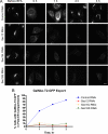
RNAi-mediated knockdown of Sec16L or Sec16S disrupts ER export. (A) ER export in RNAi-treated cells. HeLa cells stably expressing GalNAc-T2-GFP were transfected either with a control nonspecific RNAi or with an RNAi against Sec16L, Sec16S, or Sec12. At 36 h after transfection, the cells were treated with 5 μg/ml BFA for 30 min to redistribute GalNAc-T2-GFP into the ER. The cells were then washed in BFA-free medium, and at the indicated times after BFA removal, samples were fixed for fluorescence microscopy to assay for export of GalNAc-T2-GFP to post-ER compartments. All of the images in this figure were taken at the same exposure and processed in parallel. Bar, 20 μm. (B) Quantitation of the experiment shown in A. At each time point after transfection with the indicated RNAi, 100 cells were scored for the presence or absence of GalNAc-T2-GFP in post-ER compartments.
Figure 5.
Figure 5.
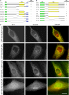
Mapping of tER localization determinants in Sec16L and Sec16S. (A) Deletion analysis of Sec16L. The starting construct was full-length Sec16L tagged at its N terminus with GFP (green box). The central conserved domain (CCD, residues 1267-1713) is shown as a yellow box, and the C-terminal conserved domain (C, residues 1928-2154) is shown as a blue box. Deletions are represented by thin angled lines. The numbers at the left indicate the residues that remained after the deletion, except that |gD(924-1227) indicates the residues that were deleted. tER localization was analyzed by transient transfection into HeLa cells followed by immunofluorescence visualization of GFP and Sec23A (see Figure 2A). A + indicates strong colocalization of the construct with Sec23A, and a − indicates little or no colocalization. (B) Deletion analysis of Sec16S. The methodology was as in part (A). Residues 271-713 make up the central conserved domain. (C) tER localization conferred by small regions of Sec16L and Sec16S. In the top two rows, GFP was fused to residues 924-1227 of Sec16Lor to residues 34-224 of Sec16S. In the bottom two rows, GFP was fused to full-length Sec16L lacking residues 924-1227 or to full-length Sec16S lacking residues 34-224. Immunofluorescence of transiently transfected cells was performed as in Figure 2A. The merged images show localization of the GFP fusion proteins (green) relative to Sec23A (red). Bar, 20 |gmm.
Figure 6.
Figure 6.
Expression of the C-terminal conserved domain of Sec16L disrupts tER and Golgi compartments. (A) Sequence alignment showing a portion of the C-terminal conserved domain of Sec16L or Sec16 from each of the indicated species. Residues matching the consensus are highlighted in yellow. (B) Expression of a fusion between GFP and the C-terminal conserved domain of Sec16L (residues 1943-2154) in HeLa cells. At 36 h after transfection, cells were subjected to immunofluorescence microscopy to visualize GFP (green) plus either Sec23A or giantin (red). In the field on the left, the single transfected cell is marked with an asterisk (*) and is the only visible cell with a disrupted Sec23A pattern. In the field on the right, the single nontransfected cell is marked with a cross-hatch (#) and is the only visible cell with a normal giantin pattern. Bar, 20 μm.
Figure 7.
Figure 7.
The C-terminal conserved domain of Sec16L interacts with Sec23A. (A) GST pull-down analysis. A GST fusion to a C-terminal fragment of Sec16L (residues 1943-2154) or Sec16S (residues 754-1060) was expressed in bacteria, and the cells were lysed and incubated with glutathione-agarose beads to bind the fusion protein. The beads were then incubated with a HeLa cell lysate. Bound proteins were eluted with glutathione and separated by SDS-PAGE, followed by immunoblotting with anti-Sec23A antibody. The input lane represents 40% of the amount of HeLa cell lysate that was used. (B) Yeast two-hybrid analysis. Gal4 fusions were expressed using a URA3“bait” vector and a LEU2“prey” vector (James et al., 1996). The C-terminal fragments used for the fusions were residues 1943-2154 of Sec16L and residues 754-1060 of Sec16S. The left panel shows an agar plate that selected for the presence of both bait and prey plasmids, whereas the other two panels show plates that also selected for two-hybrid interactions. Row 1, empty bait vector, prey vector expressing a fusion to Sec23A. Row 2, bait vector expressing a fusion to a C-terminal fragment of Sec16L or Sec16S, prey vector expressing a fusion to Sec23A. Row 3, bait vector expressing a fusion to Sec23A, prey vector expressing a fusion to a C-terminal fragment of Sec16L or Sec16S. Row 4, bait vector expressing a fusion to Sec23A, empty prey vector. Row 5, bait vector expressing a fusion to a C-terminal fragment of Sec16L or Sec16S, empty prey vector. Row 6, empty bait vector, prey vector expressing a fusion to a C-terminal fragment of Sec16L or Sec16S.
Figure 8.
Figure 8.
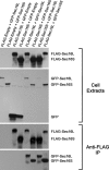
Sec16L and Sec16S are apparently present in multiple copies in a heteromeric complex. HeLa cells were transfected with a plasmid encoding either GFP alone (GFP-Empty), GFP-Sec16L, or GFP-Sec16S, and were simultaneously transfected with a second plasmid encoding either a FLAG tag alone (FLAG-Empty), FLAG-Sec16L, or FLAG-Sec16S. At 18 h after transfection, cell extracts were prepared. In the top two panels, 60 μg of soluble cell extract protein was run in each lane of an SDS-PAGE gel, and the samples were then subjected to immunoblotting with either anti-FLAG antibody (top) or anti-GFP antibody (second panel from top) to confirm that the expected proteins had been produced. In the bottom two panels, the extracts were subjected to immunoprecipitation with anti-FLAG antibody, followed by immunoblotting with either anti-FLAG antibody (second panel from bottom) or anti-GFP antibody (bottom panel). Additional lanes from the bottom two gels (data not shown) revealed that the efficiency of immunoprecipitation of the FLAG-tagged proteins was 15–20%, and the efficiency of coimmunoprecipitation of the GFP-tagged proteins was also 15–20%.

References

    1. Bannykh S. I., Balch W. E. Membrane dynamics at the endoplasmic reticulum-Golgi interface. J. Cell Biol. 1997;138:1–4. - PMC - PubMed
    1. Bevis B. J., Hammond A. T., Reinke C. A., Glick B. S. De novo formation of transitional ER sites and Golgi structures in Pichia pastoris. Nat. Cell Biol. 2002;4:750–756. - PubMed
    1. Bielli A., Haney C. J., Gabreski G., Watkins S. C., Bannykh S. I., Aridor M. Regulation of Sar1 NH2 terminus by GTP binding and hydrolysis promotes membrane deformation to control COPII vesicle fission. J. Cell Biol. 2005;171:919–924. - PMC - PubMed
    1. Connerly P. L., Esaki M., Montegna E. A., Strongin D. E., Levi S., Soderholm J., Glick B. S. Sec16 is a determinant of transitional ER organization. Curr. Biol. 2005;15:1439–1447. - PubMed
    1. Duden R., Schekman R. Insights into Golgi function through mutants in yeast and animal cells. In: Berger E. G., Roth J., editors. The Golgi Apparatus. Basel, Switzerland: Birkhäuser Verlag; 1997. pp. 219–246.

Publication types

MeSH terms

Substances

Associated data