. 2007 Mar;18(3):899-909.
doi: 10.1091/mbc.e06-06-0542. Epub 2006 Dec 27.

The Cullin3 ubiquitin ligase functions as a Nedd8-bound heterodimer

Affiliations

The Cullin3 ubiquitin ligase functions as a Nedd8-bound heterodimer

Wananit Wimuttisuk et al. Mol Biol Cell. 2007 Mar.

Abstract

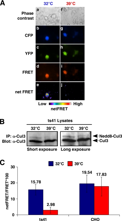
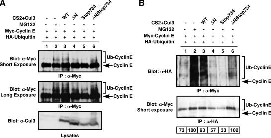
Cullins are members of a family of scaffold proteins that assemble multisubunit ubiquitin ligase complexes to confer substrate specificity for the ubiquitination pathway. Cullin3 (Cul3) forms a catalytically inactive BTB-Cul3-Rbx1 (BCR) ubiquitin ligase, which becomes functional upon covalent attachment of the ubiquitin homologue neural-precursor-cell-expressed and developmentally down regulated 8 (Nedd8) near the C terminus of Cul3. Current models suggest that Nedd8 activates cullin complexes by providing a recognition site for a ubiquitin-conjugating enzyme. Based on the following evidence, we propose that Nedd8 activates the BCR ubiquitin ligase by mediating the dimerization of Cul3. First, Cul3 is found as a neddylated heterodimer bound to a BTB domain-containing protein in vivo. Second, the formation of a Cul3 heterodimer is mediated by a Nedd8 molecule, which covalently attaches itself to one Cul3 molecule and binds to the winged-helix B domain at the C terminus of the second Cul3 molecule. Third, complementation experiments revealed that coexpression of two distinct nonfunctional Cul3 mutants can rescue the ubiquitin ligase function of the BCR complex. Likewise, a substrate of the BCR complex binds heterodimeric Cul3, suggesting that the Cul3 complex is active as a dimer. These findings not only provide insight into the architecture of the active BCR complex but also suggest assembly as a regulatory mechanism for activation of all cullin-based ubiquitin ligases.


Figures

Figure 1.
Figure 1.
Cul3–Cul3 intermolecular interactions are detected in vivo. (A) HEK293 cells expressing CFP-Cul3 (a–e), YFP-Cul3 (f–j), and CFP-Cul3 with YFP-Cul3 (k–o) were imaged for quantitative FRET analysis. Phase contrast (a, f, and k), CFP (b, g, and l), YFP (c, h, and m), and FRET fluorescence images (d, i, and n) were acquired and net FRET values (e, j, and o) were determined using the MetaMorph application. (B) Normalized net FRET values of full-length Cul3 in HEK293 cells expressing CFP–Cul3 (left), YFP–Cul3 (middle), and CFP–Cul3 with YFP–Cul3 (right). (C) HEK293 cells were transfected with vectors expressing FLAG-tagged and Myc-tagged Cul3 or a non-BTB binding mutant (L52AE55A) as indicated. Lysates were prepared, checked for protein expression (bottom), and immunoprecipitated with anti-Myc antibody. The precipitates were separated by SDS-PAGE and immunoblotted with anti-FLAG antibody (top). The upper band of the Cul3 doublet represents the Nedd8-conjugated form of Cul3. (D) FLAG-tagged Cul3 and Myc-tagged Cul3, Cul3K712R mutant, Cul3patch A mutant (A705D A706D I707E V708E), Cul3patch B mutant (P738E V739E V740D I741D), Cul3K712Rpatch B mutant, or Cul3patch A patch B mutant were expressed in HEK293 cells (middle). Lysates were prepared, and proteins were subjected to immunoprecipitation with anti-Myc antibody and immunoblotting with anti-FLAG antibody (top). Relative locations of the Cul3 mutants are illustrated by sequence alignment of human Cul1, Cul2, and Cul3 along with the predicted secondary structure of the WH-B domain (bottom).
Figure 2.
Figure 2.
The neddylation pathway is essential for Cul3–Cul3 interactions. (A) Ts41 cells expressing CFP–Cul3 and YFP–Cul3 proteins were used for quantitative FRET imaging. Cells were incubated at permissive temperature (32°C) (a–e) and nonpermissive temperature (39°C) (f–j) for 16 h before the FRET measurement. The net FRET values were calculated using MetaMorph (e and j). (B) Ts41 cell lysates at permissive (32°C) and nonpermissive (39°C) temperature were immunoprecipitated with anti-Cul3 antibody. The precipitates were separated by SDS-PAGE and immunoblotted with anti-Cul3 antibody. (C) The net FRET values from ts41 cells and CHO cells expressing CFP–Cul3 and YFP–Cul3 were normalized, and data from permissive (32°C) (blue) and nonpermissive (39°C) (red) temperatures were compared.
Figure 3.
Figure 3.
Nedd8 mediates the formation of Cul3 heterodimers by interacting with the winged-helix B domain near the C termini of Cul3. (A) HEK293 cells were transfected with HA-tagged Nedd8 (left) or harvested without transfection (right). Cell lysates were immunoprecipitated with anti-Cul3 (left, lanes 1 and 3; right, lane 1), anti-HA (left, lanes 2 and 4), or anti-Nedd8 (right, lane 2) antibodies. The precipitates were separated by SDS-PAGE and immunoblotted with anti-Cul3 (left, lanes 1 and 2; right) and anti-HA (left, lanes 3 and 4) antibodies, respectively. The intensity of Cul3 bands was quantified, and the highest value from each lane was normalized to 100% (bottom). (B) HEK293 cells expressing FLAG-tagged Cul3 and His-tagged Nedd8 were harvested. Cell lysates were prepared and the FLAG-tagged Cul3 complex was purified by precipitating via His-tagged Nedd8. Eluants were loaded onto a gel filtration column, and fractions were collected, acetone precipitated, and immunoblotted with anti-FLAG antibody (top). His-tagged Cul3 protein encoded on the pET16Cul3 vector was expressed via IPTG induction in BL21Gold (DE3) pLysS E. coli. Cells were lysed and the His-tagged Cul3 monomer was purified. The Cul3 monomer was loaded onto a gel filtration column and fractions were collected and immunoblotted with anti-Cul3 antibody (bottom). (C) HEK293 cells were transfected with vectors expressing FLAG-tagged Cul3, FLAG-tagged Cul3 mutants, and HA-tagged Nedd8 as indicated. Proteins were immunoblotted with anti-FLAG antibody (bottom) to verify expression. Cell lysates were subjected to immunoprecipitation with anti-HA antibody followed by immunoblotting with anti-FLAG antibody (top). (D) Proposed model for the formation of Cul3 heterodimers. The Cul3 dimer is held together by Nedd8 through both an isopeptide linkage and hydrophobic interactions to the Nedd8 surface.
Figure 4.
Figure 4.
The Cul3 heterodimer can bind a BTB domain-containing protein. (A) HEK293 cells were transfected with vectors encoding HA-tagged Nedd8, Myc-tagged SPOP, FLAG-tagged Cul3, and a non-BTB-binding Cul3 mutant (L52AE55A) as indicated. Protein expression was shown by immunoblotting with anti-FLAG or anti-Myc antibodies (bottom two panels). Cell lysates were subjected to immunoprecipitation with anti-FLAG (middle) or anti-HA antibodies (top) followed by immunoblotting with anti-Myc antibody. (B) HEK293 cells were transfected with vectors encoding Myc-tagged-SPOP, His-tagged Cul3, FLAG-tagged Cul3, and a non-BTB binding Cul3 mutant (L52AE55A). Protein expression was shown by immunoblotting with anti-FLAG, anti-His, and anti-Myc antibodies (bottom three panels). Cell lysates were subjected to immunoprecipitation with anti-FLAG antibody and immunoblotting with anti-Myc antibody (top).
Figure 5.
Figure 5.
A Cul3 substrate, cyclin E, binds Cul3 in a heterodimeric form. FLAG-tagged Cul3 expressed in HEK293 cells was immunoblotted with anti-FLAG antibody (bottom). Cell lysates were subjected to immunoprecipitation with anti-FLAG (lane 1) or anti-cyclin E antibodies (lane 2) followed by immunoblotting with anti-FLAG antibody (top).
Figure 6.
Figure 6.
The Cul3 heterodimer is an active form of the Cul3-based ubiquitin ligase complex. (A) HEK293 cells were transiently transfected with vectors encoding untagged Cul3, N-terminally truncated Cul3 mutant (ΔN), C-terminally truncated Cul3 mutant (stop734), Myc-tagged cyclin E, and HA-tagged ubiquitin as indicated. The proteasome inhibitor MG132 was added to the transfected cells in lane 2 18 h before harvesting. Total Cul3 protein expression from the lysates is shown (bottom). Relative levels of cyclin E ubiquitination were determined by immunoprecipitation and immunoblotting with anti-Myc antibody (top and middle). (B) The immunoblot from A was then stripped and reprobed with anti-HA antibody (top). The same lysates were also immunoprecipitated with anti-HA antibody and immunoblotted with anti-Myc antibody to determine the relative levels of ubiquitinated cyclin E in each lane (middle). Arrow denotes the position of nonubiquitinated cyclin E. The amount of ubiquitinated cyclin E was quantified relative to the amount in lane 2, which was normalized to 100% (bottom).
Figure 7.
Figure 7.
Proposed models for an active Cul3 heterodimer. A cartoon representation of the Cul3 heterodimer is illustrated based on the predicted Cul3 structure and the published Nedd8 crystal structure, showing that the formation of the Cul3 heterodimer positions the E2 binding site of one Cul3 molecule (green) in proximity to the binding site of a BTB domain-containing protein of the other Cul3 molecule (red). A single Nedd8 (yellow) is held between the two Cul3 molecules by a pair of WH-B domains. (A) The Cul3 dimer in a head-to-head arrangement. (B) The Cul3 dimer in a head-to-tail arrangement.

Similar articles

Cited by

References

    1. Angers S., Thorpe C. J., Biechele T. L., Goldenberg S. J., Zheng N., MacCoss M. J., Moon R. T. The KLHL12-Cullin-3 ubiquitin ligase negatively regulates the Wnt-beta-catenin pathway by targeting Dishevelled for degradation. Nat. Cell Biol. 2006;8:348–357. - PubMed
    1. Ciechanover A., Heller H., Elias S., Haas A. L., Hershko A. ATP-dependent conjugation of reticulocyte proteins with the polypeptide required for protein degradation. Proc. Natl. Acad. Sci. USA. 1980;77:1365–1368. - PMC - PubMed
    1. Clurman B. E., Sheaff R. J., Thress K., Groudine M., Roberts J. M. Turnover of cyclin E by the ubiquitin-proteasome pathway is regulated by cdk2 binding and cyclin phosphorylation. Genes Dev. 1996;10:1979–1990. - PubMed
    1. Cope G. A., Suh G. S., Aravind L., Schwarz S. E., Zipursky S. L., Koonin E. V., Deshaies R. J. Role of predicted metalloprotease motif of Jab1/Csn5 in cleavage of Nedd8 from Cul1. Science. 2002;298:608–611. - PubMed
    1. Geyer R., Wee S., Anderson S., Yates J., Wolf D. A. BTB/POZ domain proteins are putative substrate adaptors for Cullin 3 ubiquitin ligases. Mol. Cell. 2003;12:783–790. - PubMed

Publication types

LinkOut - more resources