

An oculomotor decision process revealed by functional magnetic resonance imaging
- PMID: 17192434
- PMCID: PMC6674715
- DOI: 10.1523/JNEUROSCI.4243-06.2006
An oculomotor decision process revealed by functional magnetic resonance imaging
Abstract
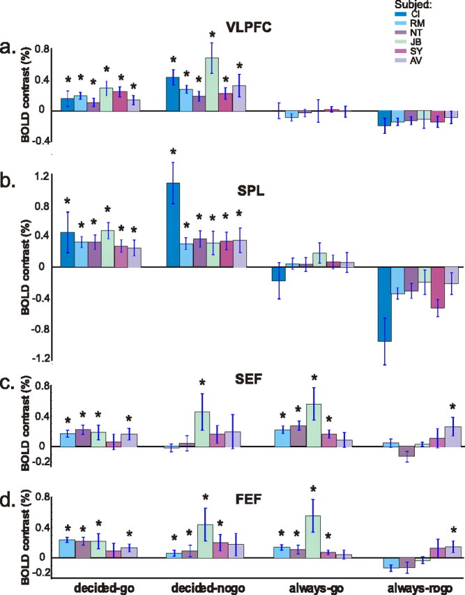
It is not known how the brain decides to act on moving objects. We demonstrated previously that neurons in the macaque supplementary eye field (SEF) reflect the rule of ocular baseball, a go/nogo task in which eye movements signal the rule-guided interpretation of the trajectory of a target. In ocular baseball, subjects must decide whether to pursue a moving spot target with an eye movement after discriminating whether the target will cross a distal, visible line segment. Here we identify cortical regions active during the ocular baseball task using event-related human functional magnetic resonance imaging (fMRI) and concurrent eye-movement monitoring. Task-related activity was observed in the SEF, the frontal eye field (FEF), the superior parietal lobule (SPL), and the right ventrolateral prefrontal cortex (VLPFC). The SPL and right VLPFC showed heightened activity only during ocular baseball, despite identical stimuli and oculomotor demands in the control task, implicating these areas in the decision process. Furthermore, the right VLPFC but not the SPL showed the greatest activation during the nogo decision trials. This suggests both a functional dissociation between these areas and a role for the right VLPFC in rule-guided inhibition of behavior. In the SEF and FEF, activity was similar for ocular baseball and a control eye-movement task. We propose that, although the SEF reflects the ocular baseball rule, both areas in humans are functionally closer to motor processing than the SPL and the right VLPFC. By recording population activity with fMRI during the ocular baseball task, we have revealed the cortical substrate of an oculomotor decision process.
Figures


Similar articles
-
Contrasting the roles of the supplementary and frontal eye fields in ocular decision making.J Neurophysiol. 2014 Jun 15;111(12):2644-55. doi: 10.1152/jn.00543.2013. Epub 2014 Mar 26. J Neurophysiol. 2014. PMID: 24671543 Free PMC article.
-
Trajectory interpretation by supplementary eye field neurons during ocular baseball.J Neurophysiol. 2005 Aug;94(2):1385-91. doi: 10.1152/jn.00109.2005. Epub 2005 May 11. J Neurophysiol. 2005. PMID: 15888531
-
Direction-dependent visual cortex activation during horizontal optokinetic stimulation (fMRI study).Hum Brain Mapp. 2006 Apr;27(4):296-305. doi: 10.1002/hbm.20185. Hum Brain Mapp. 2006. PMID: 16080162 Free PMC article.
-
The role of supplementary eye field in goal-directed behavior.J Physiol Paris. 2015 Feb-Jun;109(1-3):118-28. doi: 10.1016/j.jphysparis.2015.02.002. Epub 2015 Feb 23. J Physiol Paris. 2015. PMID: 25720602 Free PMC article. Review.
-
[Parietal Association Area and Motion Information Processing].Brain Nerve. 2016 Nov;68(11):1335-1343. doi: 10.11477/mf.1416200597. Brain Nerve. 2016. PMID: 27852024 Review. Japanese.
Cited by
-
Common and differential ventrolateral prefrontal activity during inhibition of hand and eye movements.J Neurosci. 2007 Sep 12;27(37):9893-900. doi: 10.1523/JNEUROSCI.2837-07.2007. J Neurosci. 2007. PMID: 17855604 Free PMC article.
-
Flexible interpretation of a decision rule by supplementary eye field neurons.J Neurophysiol. 2011 Dec;106(6):2992-3000. doi: 10.1152/jn.01134.2010. Epub 2011 Sep 7. J Neurophysiol. 2011. PMID: 21900513 Free PMC article.
-
Triangulating a cognitive control network using diffusion-weighted magnetic resonance imaging (MRI) and functional MRI.J Neurosci. 2007 Apr 4;27(14):3743-52. doi: 10.1523/JNEUROSCI.0519-07.2007. J Neurosci. 2007. PMID: 17409238 Free PMC article.
-
Sensorimotor-independent prefrontal activity during response inhibition.Hum Brain Mapp. 2014 May;35(5):2119-36. doi: 10.1002/hbm.22315. Epub 2013 Jun 25. Hum Brain Mapp. 2014. PMID: 23798325 Free PMC article.
-
Rapid-response impulsivity: definitions, measurement issues, and clinical implications.Personal Disord. 2015 Apr;6(2):168-181. doi: 10.1037/per0000100. Personal Disord. 2015. PMID: 25867840 Free PMC article. Review.
References
-
- Albright TD. Direction and orientation selectivity of neurons in visual area MT of the macaque. J Neurophysiol. 1984;52:1106–1130. - PubMed
-
- Amador N, Schlag-Rey M, Schlag J. Primate antisaccade. II. Supplementary eye field neuronal activity predicts correct performance. J Neurophysiol. 2004;91:1672–1689. - PubMed
-
- Aron AR, Fletcher PC, Bullmore ET, Sahakian BJ, Robbins TW. Stop-signal inhibition disrupted by damage to right inferior frontal gyrus in humans. Nat Neurosci. 2003;6:115–116. - PubMed
-
- Baddeley A, Della Sala S. Working memory and executive control. In: Roberts AC, Robbins TW, Weiskrantz L, editors. The prefrontal cortex: executive and cognitive functions. New York: Oxford UP; 1998. pp. 9–21.
-
- Bandettini PA, Jesmanowicz A, Wong EC, Hyde JS. Processing strategies for time-course data sets in functional MRI of the human brain. Magn Reson Med. 1993;30:161–173. - PubMed
Publication types
MeSH terms
Grants and funding
LinkOut - more resources
Full Text Sources
Medical