Case Reports
. 2006 Dec 28;355(26):2757-64.
doi: 10.1056/NEJMoa063804.

Deficiency of cartilage-associated protein in recessive lethal osteogenesis imperfecta

Affiliations
Case Reports

Deficiency of cartilage-associated protein in recessive lethal osteogenesis imperfecta

Aileen M Barnes et al. N Engl J Med. .

Abstract

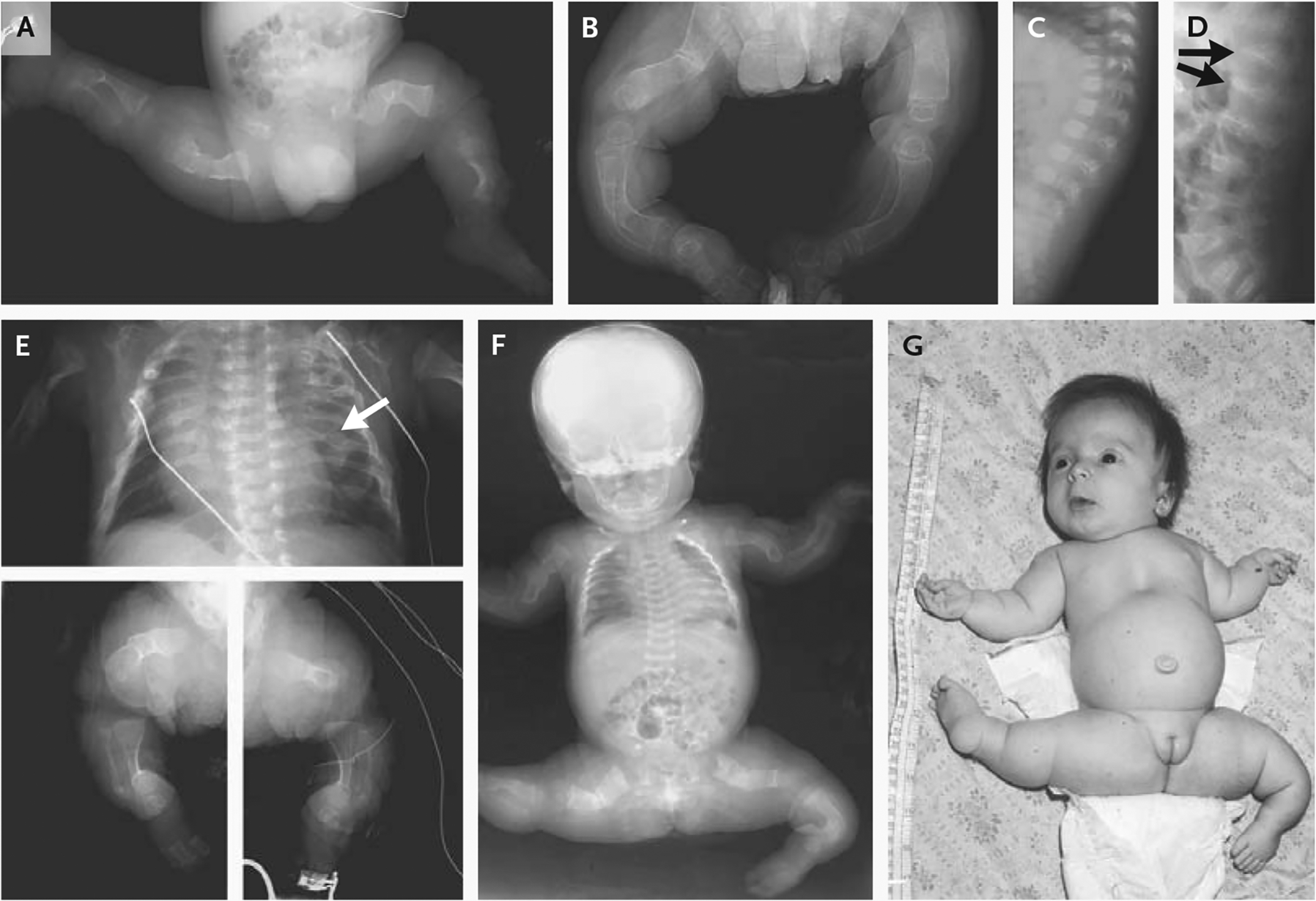
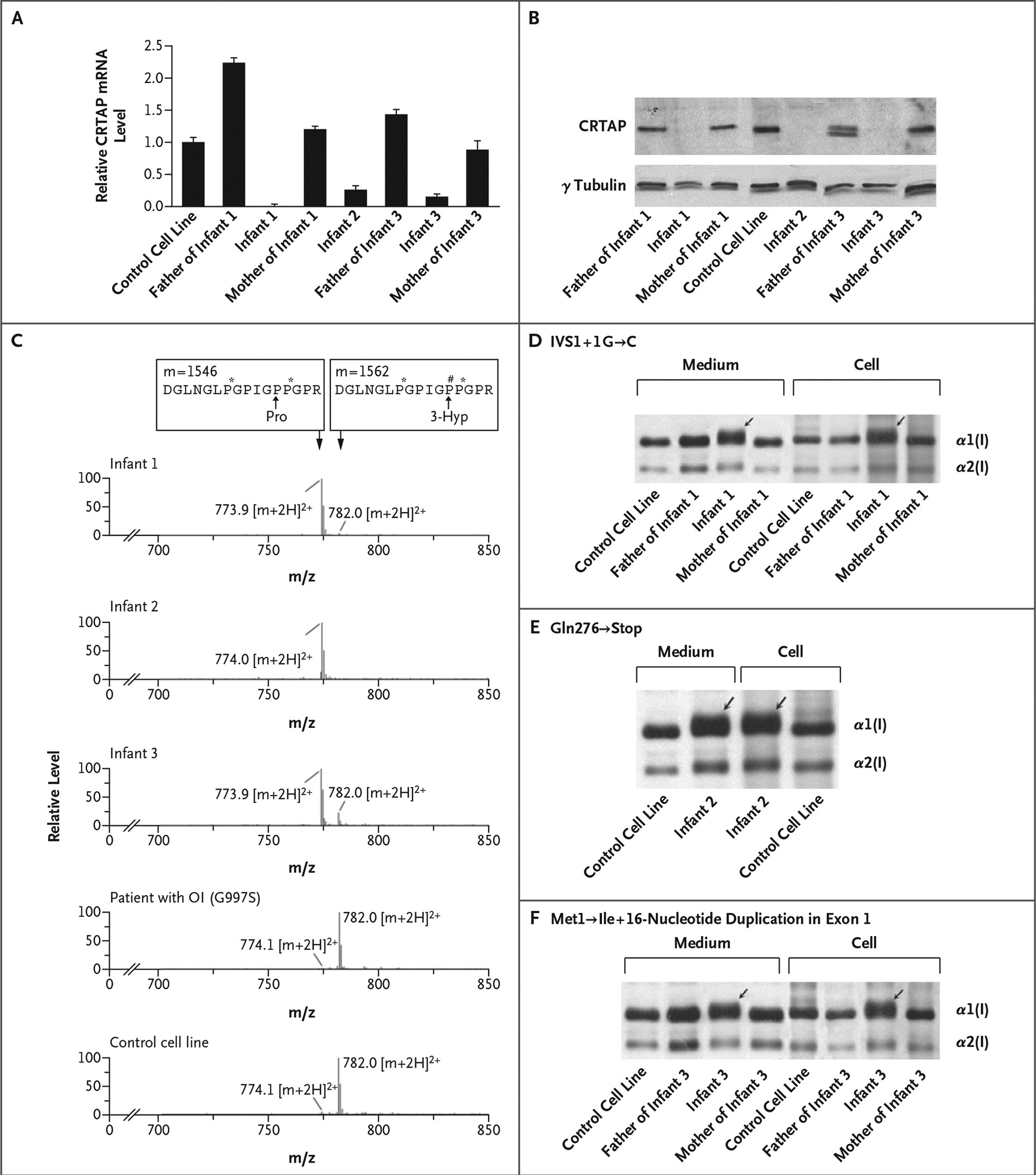
Classic osteogenesis imperfecta, an autosomal dominant disorder associated with osteoporosis and bone fragility, is caused by mutations in the genes for type I collagen. A recessive form of the disorder has long been suspected. Since the loss of cartilage-associated protein (CRTAP), which is required for post-translational prolyl 3-hydroxylation of collagen, causes severe osteoporosis in mice, we investigated whether CRTAP deficiency is associated with recessive osteogenesis imperfecta. Three of 10 children with lethal or severe osteogenesis imperfecta, who did not have a primary collagen defect yet had excess post-translational modification of collagen, were found to have a recessive condition resulting in CRTAP deficiency, suggesting that prolyl 3-hydroxylation of type I collagen is important for bone formation.


Conflict of interest statement

No potential conflict of interest relevant to this article was reported.

Figures

Figure 1.
Figure 1.. Clinical Characteristics of Infants with CRTAP Deficiency.
Panels A and B show the legs of Infant 1 at birth and at 8 months of age, respectively. His long bones were severely osteopenic and crumpled at birth; at 8 months, most fractures had healed but the lack of modeling (undertubulation) was indicated by the cylindrical configuration of diaphyses. Panels C and D show lateral views of the spine of Infant 1 at birth and 8 months of age, respectively. The vertebrae were severely osteoporotic at birth but had a normal geometry; by 8 months, there were multiple vertebral compressions (arrows), especially at vertebrae T12 and L1. Panel E shows a composite of radiographs of Infant 2 at birth. The chest was narrow, and the cardiac silhouette was enlarged. Multiple beads of callus (arrow) represent sites of healing fractures. Long bones in the legs were extremely osteopenic and crumpled, with poor modeling and a cylindrical configuration. Panel F shows a radiograph and Panel G a photograph of Infant 3 at 2 months of age. The thorax was narrow, with multiple healing rib fractures and extremely osteoporotic long bones showing crumpled, poorly modeled geometry. Infant 3 held her legs in an abducted position, which is typical of osteogenesis imperfecta; in contrast to infants with osteogenesis imperfecta type II, she had a round face (rather than triangular), a normal profile (rather than a flat midface), and a long philtrum.
Figure 2.
Figure 2.. Results of Analyses of CRTAP mRNA and Biochemical Analyses of CRTAP and Type I Collagen.
Panel A shows the relative expression of CRTAP in fibroblasts, measured using real-time RT-PCR as the CRTAP mRNA level normalized to the glyceraldehyde-3-phosphate dehydrogenase (GAPDH) mRNA level and then to the CRTAP mRNA level in the control fibroblast line (which was arbitrarily set at 1). T bars represent standard deviations. Panel B shows a Western blot of fibroblast CRTAP, which was lacking in the affected infants and present in their parents. The father of Infant 3 also had a truncated form of CRTAP. Panel C shows the results of tandem mass spectrometry, revealing a low level of Pro986 3-hydroxylation or none in Infants 1, 2, and 3, in contrast to the normal Pro986 3-hydroxylation in both a patient with classic dominant osteogenesis imperfecta (OI), caused by a glycine substitution (Gly997→Ser) in collagen, and in a control cell line. The tracing shows the cumulative, full scan across the liquid chromatography peak. The top boxes represent the masses (m, in atomic mass units) and the amino acid sequences (capital letters; P# and P* denote 3- and 4-hydroxyproline [Hyp], respectively) of collagen peptides that have either undergone 3-hydroxylation at the proline at position 986 (Pro986) (right-hand box, in which the proline is converted to 3-Hyp) or have not (left-hand box). The levels of the ions relative to one another are shown, as are their mass:charge (m/z) ratios; shown are the double-charged peptide ions ([m+2H]2+) that differ by the 16 mass units of an additional oxygen atom. Peptide ions that have undergone normal Pro986 hydroxylation have an m/z ratio of 782.0, whereas peptides that have not have an m/z ratio of approximately 774. The results of SDS-urea-PAGE of type I collagen from the three infants, their parents, and the control cell line are shown; Infant 1 had a IVS1+1G→C substitution (Panel D); Infant 2, a Gln276→ Stop substitution (Panel E); and Infant 3, a Met1→Ile substitution and a 16-nucleotide duplication in exon 1 (Panel F). The broad bands representing the α1(I) chains indicate the presence of a form with delayed migration in Infants 1, 2, and 3 that was also secreted into the culture medium (arrows).

Similar articles

Cited by

References

    1. Marini JC. Osteogenesis imperfecta In: Behrman RE, Kliegman RM, Jenson HB, eds. Nelson textbook of pediatrics. 17th ed. Philadelphia: W.B. Saunders, 2004: 2336–8.
    1. Sillence DO, Senn A, Danks DM. Genetic heterogeneity in osteogenesis imperfecta. J Med Genet 1979;16:101–16. - PMC - PubMed
    1. Byers P, Cole W. Osteogenesis imperfecta In: Royce PM, Steinmann B, eds. Connective tissue and its heritable disorders: molecular, genetic, and medical aspects. 2nd ed. New York: Wiley-Liss, 2002: 385–430.
    1. Bruckner P, Eikenberry EF. Formation of the triple helix of type I procollagen in cellulo: temperature-dependent kinetics support a model based on cis in equilibrium trans isomerization of peptide bonds. Eur J Biochem 1984;140:391–5. - PubMed
    1. Aitchison K, Ogilvie D, Honeyman M, Thompson E, Sykes B. Homozygous osteogenesis imperfecta unlinked to collagen I genes. Hum Genet 1988;78:233–6. - PubMed

Publication types