. 2007 Jan;16(1):52-68.
doi: 10.1110/ps.062444807.

Contributions of the interdomain loop, amino terminus, and subunit interface to the ligand-facilitated dimerization of neurophysin: crystal structures and mutation studies of bovine neurophysin-I

Affiliations

Contributions of the interdomain loop, amino terminus, and subunit interface to the ligand-facilitated dimerization of neurophysin: crystal structures and mutation studies of bovine neurophysin-I

Xintian Li et al. Protein Sci. 2007 Jan.

Abstract

Current evidence indicates that the ligand-facilitated dimerization of neurophysin is mediated in part by dimerization-induced changes at the hormone binding site of the unliganded state that increase ligand affinity. To elucidate other contributory factors, we investigated the potential role of neurophysin's short interdomain loop (residues 55-59), particularly the effects of loop residue mutation and of deleting amino-terminal residues 1-6, which interact with the loop and adjacent residues 53-54. The neurophysin studied was bovine neurophysin-I, necessitating determination of the crystal structures of des 1-6 bovine neurophysin-I in unliganded and liganded dimeric states, as well as the structure of its liganded Q58V mutant, in which peptide was bound with unexpectedly increased affinity. Increases in dimerization constant associated with selected loop residue mutations and with deletion of residues 1-6, together with structural data, provided evidence that dimerization of unliganded neurophysin-I is constrained by hydrogen bonding of the side chains of Gln58, Ser56, and Gln55 and by amino terminus interactions, loss or alteration of these hydrogen bonds, and probable loss of amino terminus interactions, contributing to the increased dimerization of the liganded state. An additional intersubunit hydrogen bond from residue 81, present only in the liganded state, was demonstrated as the largest single effect of ligand binding directly on the subunit interface. Comparison of bovine neurophysins I and II indicates broadly similar mechanisms for both, with the exception in neurophysin II of the absence of Gln55 side chain hydrogen bonds in the unliganded state and a more firmly established loss of amino terminus interactions in the liganded state. Evidence is presented that loop status modulates dimerization via long-range effects on neurophysin conformation involving neighboring Phe22 as a key intermediary.


Figures

Figure 1.
Figure 1.
Amino acid sequences of BNPI and BNPII. With the exception of the F91 STOP mutants, our BNPI constructs terminate at Ser92 (Eubanks et al. 1999). Interdomain loop residues 55–59 are shown in bold. Residues at the subunit interface in the unliganded state are underlined. Binding site residues are italicized. Note that residue 82 is not an interface residue, although inadvertently listed as such in an earlier reference (Naik et al. 2005).
Figure 2.
Figure 2.
Scatchard plots of binding the peptide phenylalanyl-tyrosine amide to WT BNPI (open circles), the single mutants Q58V (filled triangles) and Q55A (filled squares), and the double mutant Q58V/Q55A (filled circles). Ordinate units are inverse molarities (M−1). Conditions: ∼0.1 mM protein, pH 6.2, 25°C. Binding constants in units of inverse molarity for these specific experiments calculated from the data as described in Materials and Methods are: Q55A (2480), WT (14,860), Q58V/Q55A (17,780), and Q58V (64,400). Note the curvature of the data obtained at values of formula image <0.5 for the double mutant as described in Materials and Methods, which, in this case, would not have significantly influenced the calculated binding constant.
Figure 3.
Figure 3.
NMR spectra of WT BNPI and its des 1–6 and desQ58V mutants at ∼0.2 mM concentration illustrating the determination of dimerization constants. Signals listed as M and D are the monomer and dimer signals respectively of the α-proton signal of Cys28. The signal marked with an x is assigned as noise or a trace contaminant based on its absence in other spectra of the same proteins and is not included in calculations. (Top) WT BNPI, 0.23 mM, M/D = 1.01 ± 0.08, calculated dimerization K = 4350 M−1. (Middle) des 1–6 BNPI, 0.16 mM, M/D = 0.84 ± 0.01, calculated dimerization K = 8200 M−1. (Bottom) desQ58V, 0.23 mM, M/D = 0.425, calculated dimerization K = 17,950 M−1. Note that calculated dimerization constants apply only to the individual spectra shown and variably deviate to some degree from the composite results reported in ▶.
Figure 4.
Figure 4.
Effect of binding the dipeptide FF to the hormone-binding site of the des 1–6 Q58V mutant of BNPI as monitored by 1D proton NMR. The chemical shifts of Cys28 α-proton signals in monomeric (M) and dimeric (D) states are indicated; note that these are essentially independent of the state of protein ligation (for example, see Barat et al. 2004). FF is chosen for NMR binding studies because its signals in free and bound states do not obscure the Cys28 signals; the binding constant of FF is similar to that of FY. (Lower spectrum) 0.1 mM protein, pH 7. (Upper spectrum) Same sample after addition of 0.5 mM FF. The protein is saturated with peptide under these conditions. Note the complete loss of both the Cys28 monomer signal and the 6.6 ppm signal in the presence of peptide.
Figure 5.
Figure 5.
Asymmetric units of liganded and unliganded derivatives of BNPI. (Left) des BNPIfy. The bound dipeptides are shown as stick structures. (Right) desBNPIF91STOP in the absence of peptide. Lower and upper dimers for both structures are AB (yellow and green chains) and CD (blue and orange chains), respectively. E subunits are shown in light purple. Structures were visualized by PyMOL (http://www.pymol.org).
Figure 6.
Figure 6.
Comparison of the backbone structure of desBNPIfy (black) with that of WT BNPIIfy (white) and desBNPIF91STOP (white). Positions of the interdomain loop and amino (N) and carboxyl (C) termini are indicated. Residues 1–6 of BNPIIfy were deleted from the comparison to allow the superposition. (Left) Superposition of desBNPIfy and BNPIIfy. (Right) Superposition of desBNPIfy and desBNPIF91STOP.
Figure 7.
Figure 7.
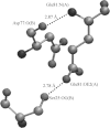
Hydrogen bonding of residues 25, 77, and 81 across the subunit interface of the AB dimer in the liganded state. Dashed lines indicate hydrogen bonds. Subunit identity is given in parentheses.
Figure 8.
Figure 8.
Superficial similarity between the crystal structures of the FY complexes of desBNPI and its Q58V mutant. (Top) Region (residues 50–61) surrounding residue 58 in the WT (black) and mutant (gray) proteins. Chain runs from left to right. Note similar side chain positions in the two proteins for residue 58 and apparent similarity in backbone structure. (Below) RMSD comparison of the A subunit structures of the two proteins. Ordinate is Ångstrom values for backbone residues (diamonds) and side chains (squares). RMSD values of 0 for some side chains indicates Gly residues.
Figure 9.
Figure 9.
Evidence that the 6.6 ppm signal of Q58 mutants is from Tyr49. (Top) DQF-COSY spectrum of the Q58V mutant. (Bottom panel, spectra from top to bottom), 1D proton spectra of Q58V mutant; WT protein; double mutant des 1–6,Q58V; and Q58V nitrated at Tyr49. (Nitration moves Tyr49 ring protons downfield from 7 ppm to the Phe ring proton region, which is not shown in the 1D spectra.) In the DQF-COSY spectrum, the principal signal from the 3,5 ring protons of Tyr49 is located at ∼6.75 ppm and shows connectivity solely to the 2,6 ring protons located at 7.13 ppm. The 6.6 ppm signal shows connectivity only to 7.09 ppm, the latter representing the 2,6 protons of the same ring also upfield shifted relative to the WT protein. Protons downfield from the Tyr49 protons in the COSY spectrum are from Phe residues and show no connectivity to the 6.6 ppm signal. Signals at ∼6.4 and 6.15 ppm are the dimer and monomer signals, respectively, of the Cys28 α-proton.
Figure 10.
Figure 10.
Representative NMR spectra showing the effect of the des 1–6 mutation on the WT protein and the effect of the Q55A mutation on the Q58V mutant. (Top to bottom) WT BNPI; des 1–6 BNPI; Q58V mutant of BNPI; and the double mutant Q55A/Q58V. Arrow points to 6.62 ppm.
Figure 11.
Figure 11.
Effect of binding ligand on the environment and interactions of the Gln58 carboxamide in des 1–6 BNPI. (Left) The unbound state showing proximity of the Gln58 carboxamide to the ring of Phe 22 in the protein interior, and hydrogen bonding of the carboxamide to the O of Gly57, and the –OH of Ser56. (Right) The bound state showing movement of the Gln58 carboxamide away from Phe22 and the loss of its hydrogen-bonding interactions. In the bound state, the distance between the Gln58 NE2 and Gly57O has increased to 5.46Å and the distance between the 58OE1 and 56OG has increased to 11.58Å.

Similar articles

Cited by

References

    1. Barat, C., Simpson, L.-R., and Breslow, E. 2004. Properties of human vasopressin constructs: Inefficient monomer folding in the absence of copeptin as a potential contributor to diabetes insipidus. Biochemistry 43 8191–8203. - PubMed
    1. Breslow, E. and Burman, S. 1990. Molecular, thermodynamic and biological aspects of recognition and function in neurophysin-hormone systems: A model system for the analysis of protein–peptide interactions. Adv. Enzymol 63 1–67. - PubMed
    1. Breslow, E., Weis, J., and Menendez-Botet, C.J. 1973. Small peptides as analogs of oxytocin and vasopressin in their interactions with bovine neurophysin-II. Biochemistry 12 4644–4653. - PubMed
    1. Breslow, E., LaBorde, T., Bamezai, S., and Scarlata, S. 1991. Binding and fluorescence studies of the relationship between neurophysin–peptide interaction and neurophysin self-association: An allosteric system exhibiting minimal cooperativity. Biochemistry 30 7990–8000. - PubMed
    1. Breslow, E., Mishra, P.K., Huang, H.-B., and Bothner-by, A. 1992. Slowly interchanging conformers of bovine neurophysin-II in the unliganded dimeric state. Biochemistry 31 11397–11404. - PubMed

Publication types

MeSH terms

LinkOut - more resources