. 2007 Jan;16(1):99-109.
doi: 10.1110/ps.062251707.

A phosphate-binding subsite in bovine pancreatic ribonuclease A can be converted into a very efficient catalytic site

Affiliations

A phosphate-binding subsite in bovine pancreatic ribonuclease A can be converted into a very efficient catalytic site

Mohammed Moussaoui et al. Protein Sci. 2007 Jan.

Abstract

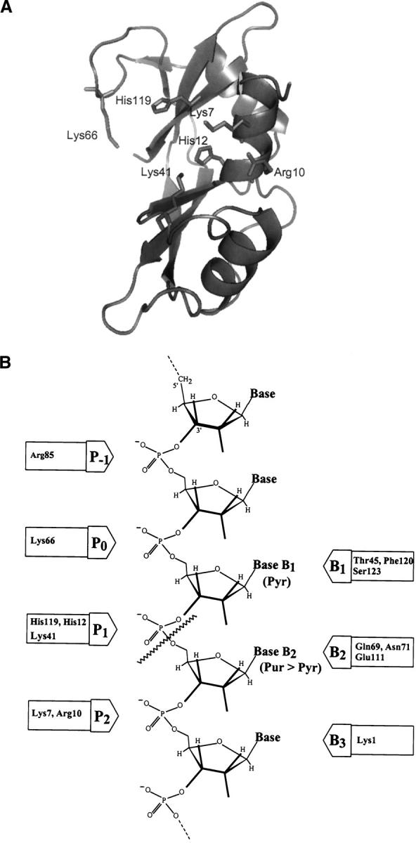
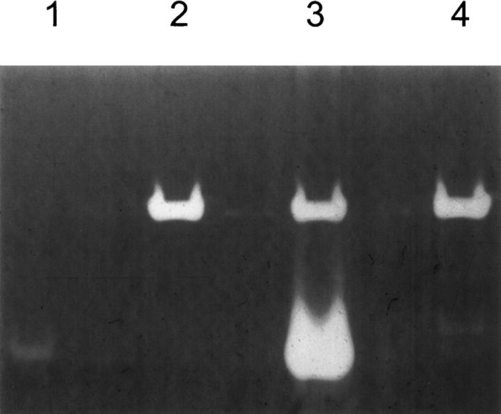
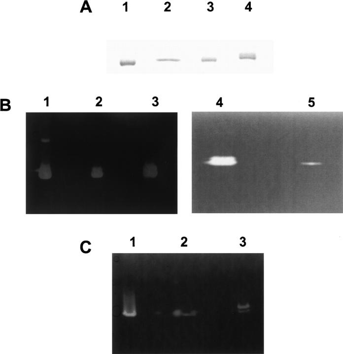
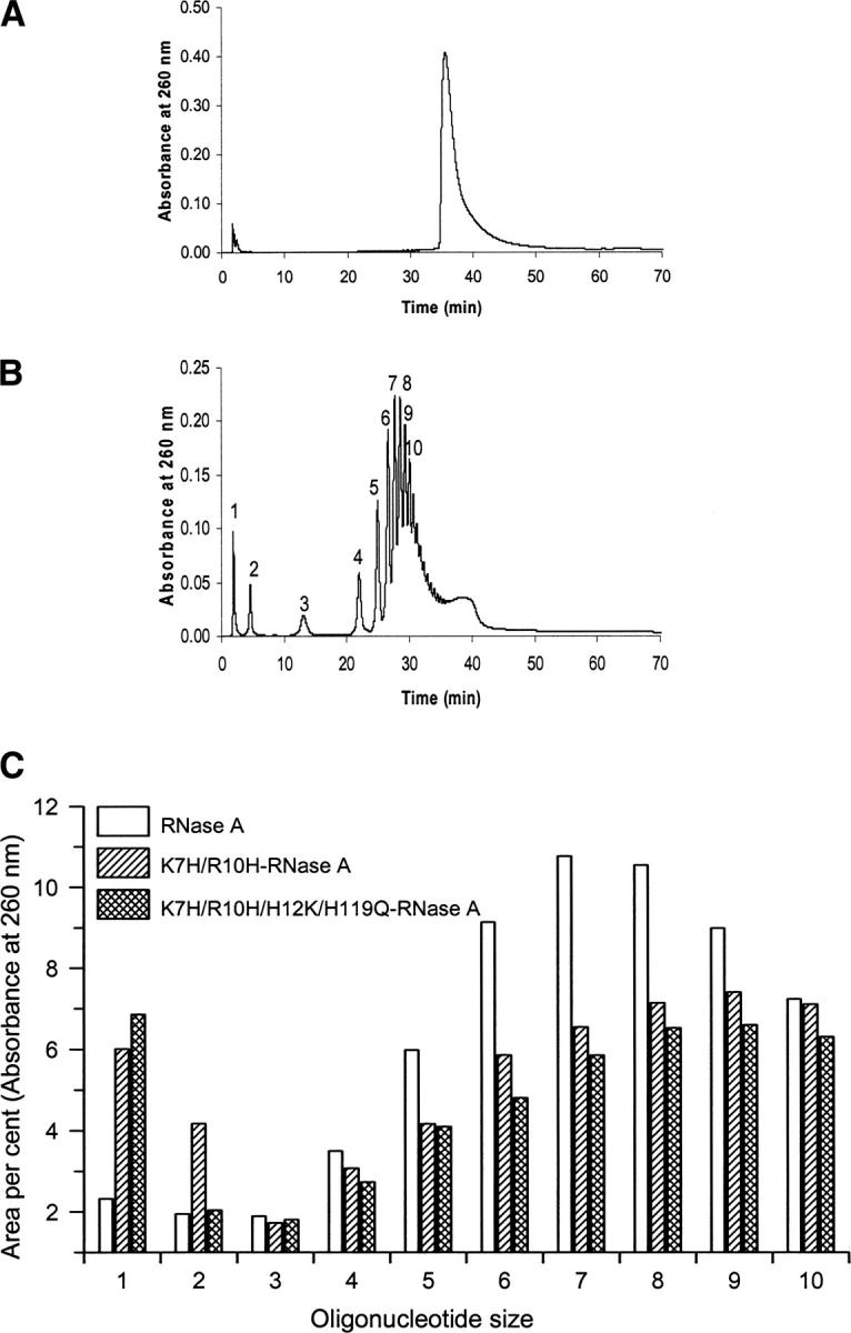
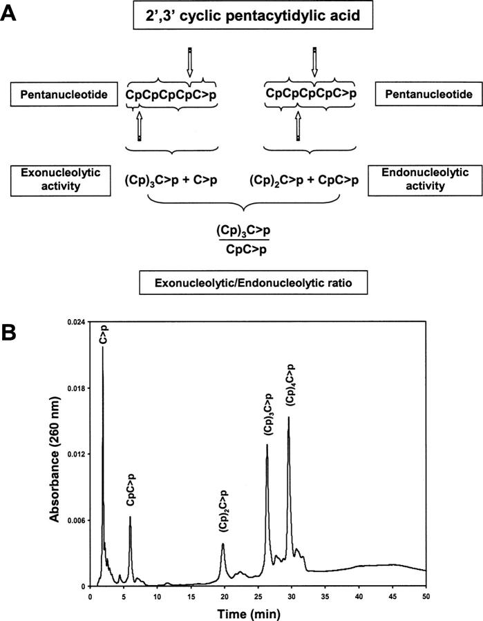
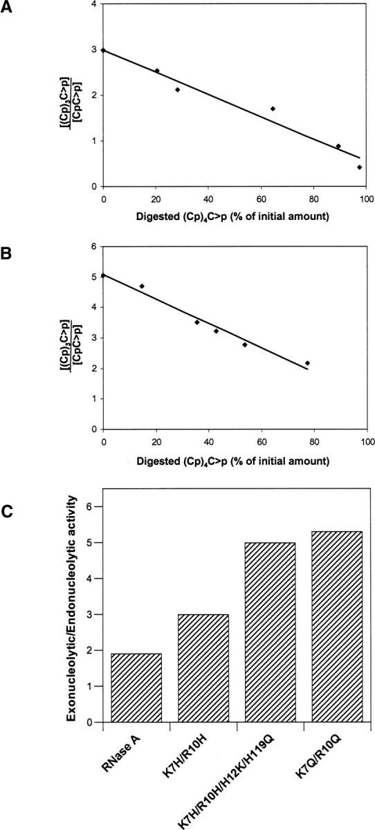
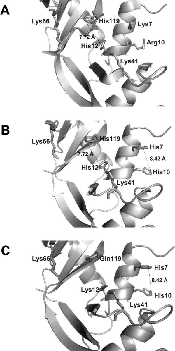
A general acid-base catalytic mechanism is responsible for the cleavage of the phosphodiester bonds of the RNA by ribonuclease A (RNase A). The main active site is formed by the amino acid residues His12, His119, and Lys41, and the process follows an endonucleolytic pattern that depends on the existence of a noncatalytic phosphate-binding subsite adjacent, on the 3'-side, to the active site; in this region the phosphate group of the substrate establishes electrostatic interactions through the side chains of Lys7 and Arg10. We have obtained, by means of site-directed mutagenesis, RNase A variants with His residues both at positions 7 and 10. These mutations have been introduced with the aim of transforming a noncatalytic binding subsite into a putative new catalytic active site. The RNase activity of these variants was determined by the zymogram technique and steady-state kinetic parameters were obtained by spectrophotometric methods. The variants showed a catalytic efficiency in the same order of magnitude as the wild-type enzyme. However, we have demonstrated in these variants important effects on the substrate's cleavage pattern. The quadruple mutant K7H/R10H/H12K/H119Q shows a clear increase of the exonucleolytic activity; in this case the original native active site has been suppressed, and, as consequence, its activity can only be associated to the new active site. In addition, the mutant K7H/R10H, with two putative active sites, also shows an increase in the exonucleolytic preference with respect to the wild type, a fact that may be correlated with the contribution of the new active site.


Figures

Figure 1.
Figure 1.
(A) Three-dimensional structure of RNase A. Amino acid residues belonging to the active site p1 (His12, His 119, and Lys41) and to the noncatalytic phosphate-binding subsites p0 (Lys66) and p2 (Lys7 and Arg10) are shown in stick. The picture was obtained using the program PyMOL (DeLano Scientific program, PDB accession code 7RSA). (B) Schematic representation of the substrate-binding sites; p1 corresponds to the active site where the cleavage of the phosphodiester bond takes place (wavy line).
Figure 2.
Figure 2.
Positive and negative controls of recombinant RNases. Activities were determined by the zymogram technique in 15% SDS-PAGE containing poly(C) as substrate. (Lane 1) 300 pg of RNase A. (Lane 2) 5 μl of the intracellular soluble fraction of E. coli cells transformed with the plasmid pET11d lacking the RNase A insert. (Lane 3) 5 μl of the intracellular soluble fraction of E. coli cells transformed with the plasmid pET11d carrying native RNase A as target gene. (Lane 4) 5 μl of the intracellular soluble fraction of E. coli cells transformed with the plasmid pET11d carrying H12K/H119Q-RNase A as target gene.
Figure 3.
Figure 3.
SDS-15% PAGE with Coomassie blue staining (A) and RNase activity staining on gels containing either poly(C) (B) or poly(U) (C) as substrates. (A) (Lane 1) native RNase A. (Lane 2) H12K/H119Q-RNase A. (Lane 3) K7H/R10H/H12K/H119Q-RNase A. (Lane 4) K7H/R10H-RNase A (B) (Lane 1) native RNase A (30 ng). (Lane 2) K7H/R10H/H12K/H119Q-RNase A (30 ng). (Lane 3) K7H/R10H-RNase A (30 ng). (Lane 4) native RNase A (0.2 ng). (Lane 5) H12K/H119Q-RNase A (80 ng). (C) (Lane 1) native RNase A (30 ng). (Lane 2) K7H/R10H/H12K/H119Q-RNase A (30 ng). (Lane 3) K7H/R10H-RNase A (30 ng).
Figure 4.
Figure 4.
Analysis by reversed-phase HPLC of the poly(C) substrate before addition of enzyme (A) and of the products obtained by digestion with RNase A (B) (the oligonucleotide size of the products is indicated). (C) Comparison of oligocytidilyc acid formation ((Cp)nC>p (n = 0–10)) from the poly(C) cleavage by RNase A and the variants K7H/R10H and K7H/R10H/H112K/H119Q. Area percent for each oligonucleotide was determined from the area of the corresponding peak eluted from a reversed-phase HPLC column (Nova pak C18). Due to the differences in the cleavage pattern and the enzyme activities, the comparison has been established using as reference the moment in which 30% of the poly(C) substrate remained undigested.
Figure 5.
Figure 5.
(A) Possible distribution of products formed by the initial cleavage of (Cp)4C>p. Exonucleolytic cleavage, from either end, yields a mononucleotide plus a tetranucleotide whereas the endonucleolytic cleavage, also from either end, yields a mixture of di- and trinucleotide. The ratio between the tetranucleotide and the dinucleotide formation may be used as an indicator of the preference for the endo- or exonucleolytic cleavage. (B) Separation by reversed-phase HPLC (Nova pak C18 column) of the products obtained from the digestion of (Cp)4C>p by K7H/R10H/H12KH119Q-RNase A in 10 mM HEPES-KOH at pH 7.5. Elution was carried out as described in Material and Methods.
Figure 6.
Figure 6.
Effect of the presence of a new active site on the exonucleolytic versus endonucleolytic activity of RNase A. Tetranucleotide/dinucleotide ratio for the cleavage of the pentacytidylic acid substrate (Cp)4C>p by K7H/R10H-RNase A (A) and by the K7H/R10H/H12K/H119Q-RNase A (B). With this substrate, tetranucleotide formation indicates exonucleolytic cleavage by the enzyme whereas formation of dinucleotide indicates endonucleolytic cleavage (the trinucleotide molecule could have been used as well). Extrapolation of the ratios to zero time (0% of initial substrate digested) was determined with the program GraFit v.5 (Leatherbarrow 2001) and indicates the preference of the enzymes on the intact substrate. The ratio decreases with time because the tetranucleotide produced at the initial stages of the reaction is later used as a substrate by the enzyme. (C) Exonucleolyticversus endonucleolytic activity of RNase A and variants as determined from the ratios of the products at zero time. The value corresponding to the variant K7Q/R10Q was taken from Cuchillo et al. (2002).
Figure 7.
Figure 7.
Three-dimensional structure of the region around the RNase A active site. (A) RNase A. (B,C) Molecular modeling of the variants K7H/R10H-RNase A and K7H/R10H/H12K/H119Q-RNase A, respectively. Molecular modeling was carried out with the program Deep View/Swiss PDB Viewer and the picture was drawn using PyMOL, DeLano Scientific program (PDB accession code 7RSA).

Similar articles

Cited by

References

    1. Alonso, J., Nogués, M.V., and Cuchillo, C.M. 1986. Modification of bovine pancreatic ribonuclease A with 6-chloropurine riboside. Arch. Biochem. Biophys. 246: 681–689. - PubMed
    1. Boix, E., Nogués, M.V., Schein, C.H., Benner, S.A., and Cuchillo, C.M. 1994. Reverse transphosphorylation by ribonuclease A needs an intact p2-binding site. Point mutations at Lys-7 and Arg-10 alter the catalytic properties of the enzyme. J. Biol. Chem. 269: 2529–2534. - PubMed
    1. Boix, E., Leonidas, D.D., Nikolovski, Z., Nogués, M.V., Cuchillo, C.M., and Acharya, K.R. 1999a. Crystal structure of eosinophil cationic protein at 2.4 Å resolution. Biochemistry 38: 16794–16801. - PubMed
    1. Boix, E., Nikolovski, Z., Moiseyev, G.P., Rosenberg, H.F., Cuchillo, C.M., and Nogués, M.V. 1999b. Kinetic and product distribution analysis of human eosinophil cationic protein indicates a site arrangement that favors exonuclease-type activity. J. Biol. Chem. 274: 15605–15614. - PubMed
    1. Bravo, J., Fernández, E., Ribó, M., de Llorens, R., and Cuchillo, C.M. 1994. A versatile negative-staining ribonuclease zymogram. Anal. Biochem. 219: 82–86. - PubMed

Publication types

LinkOut - more resources