

Evaluation of equine papillomas, aural plaques, and sarcoids for the presence of Equine papillomavirus DNA and Papillomavirus antigen
- PMID: 17193879
- PMCID: PMC1635997
Evaluation of equine papillomas, aural plaques, and sarcoids for the presence of Equine papillomavirus DNA and Papillomavirus antigen
Abstract
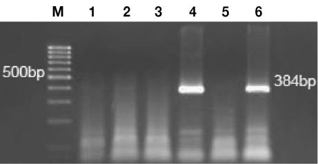
Immunohistochemical (IHC) testing and electron microscopy have implicated Papillomavirus (PV) as the etiologic agent for equine papillomas and aural plaques, but Equine papillomavirus (EPV) DNA has yet to be demonstrated in these lesions by polymerase chain reaction (PCR). The purpose of this study was to evaluate formalin-fixed, paraffin-embedded tissues from naturally occurring cases of equine papillomas, aural plaques, and sarcoids for the presence of EPV DNA by means of PCR and for the presence of PV antigen by means of IHC testing. We used EPV-specific primers that amplified a region of 384 base pairs (bp) spanning the E4 and L2 genes of the EPV genome and consensus PV primers that amplified a 102-bp region of the L1 gene. Group-specific PV structural antigens were detected with the use of a streptavidin-biotin-alkaline phosphatase IHC stain. With IHC testing, 23 of 38 papillomas, 4 of 9 aural plaques, and 0 of 10 sarcoids were positive for PV antigen; EPV DNA was found in 20 of the 38 papillomas and 1 of the 10 sarcoids but 0 of the 9 aural plaques. The consensus primers did not amplify novel PV DNA in any of the tissues. Nucleotide sequencing of viral DNA from 7 papillomas amplified with EPV-specific primers revealed DNA fragments that were 96% to 99% identical to known EPV sequences. Some samples had nucleotide substitutions in common, which suggests infection with related strains. Together, EPV DNA or PV antigen (or both) was demonstrated in 26 (68%) of the 38 equine papillomas. Although aural plaques contained PV antigen, they were negative for EPV DNA; therefore, we hypothesize that aural plaques contain a PV distinct from EPV.
Les épreuves immunohistochimiques (IHC) et la microscopie électronique ont mis en cause le Papillomavirus (PV) comme agent étiologique des papillomes équins et des plaques auriculaires (papillomes de l’oreille), mais l’ADN du papillomavirus équin (EPV) n’a pas encore été mis en évidence par réaction d’amplification en chaîne par la polymérase (PCR) dans ces lésions. Le but de l’étude était d’évaluer la présence d’ADN de l’EPV par épreuve PCR et la présence d’antigène du PV par épreuve IHC dans des tissus fixés à la formaline et paraffinés provenant de cas cliniques de papillomes équins, de plaques auriculaires et de sarcoïdes. Des amorces spécifiques à EPV amplifiant une région de 384 paires de bases (bp) couvrant les gènes E4 et L2 du génome d’EPV et des amorces consensus du PV amplifiant une région de 102 bp du gène L1 ont été utilisées. Les antigènes structuraux spécifiques de groupe du PV ont été détectés en IHC par coloration à la streptavidine–biotine–phosphatase alcaline. Par épreuve IHC, 23 des 38 papillomes, 4 des 9 plaques auriculaires et 0 des 10 sarcoïdes étaient positifs pour la présence d’antigène du PV; l’ADN d’EPV a été trouvé dans 20 des 38 papillomes, 1 des 10 sarcoïdes et 0 des 9 plaques auriculaires. Les amorces consensus n’ont pas amplifiées d’ADN nouveaux de PV dans aucun des tissus. Le séquençage nucléotidique de l’ADN viral amplifié provenant de 7 papillomes avec les amorces spécifiques à EPV a révélé des fragments d’ADN de 96 % à 99 % identiques à des séquences connues d’EPV. Quelques cas avaient des substitutions nucléotidiques en commun, ce qui suggère une infection par des souches reliées. Dans l’ensemble, l’ADN d’EPV ou l’antigène de PV (ou les deux) ont été démontrés dans 26 (68 %) des 38 papillomes équins. Bien que les plaques auriculaires contenaient de l’antigène de PV, elles se sont avérées négatives pour la présence d’ADN d’EPV; ainsi, l’hypothèse est émise que les plaques auriculaires contiennent un PV distinct de l’EPV.
(Traduit par Docteur Serge Messier)
Figures
Similar articles
-
Detection of papillomavirus-DNA in mesenchymal tumour cells and not in the hyperplastic epithelium of feline sarcoids.Vet Dermatol. 2003 Feb;14(1):47-56. doi: 10.1046/j.1365-3164.2003.00324.x. Vet Dermatol. 2003. PMID: 12603685
-
Generalized papillomatosis in three horses associated with a novel equine papillomavirus (EcPV8).Vet Dermatol. 2018 Feb;29(1):72-e30. doi: 10.1111/vde.12481. Epub 2017 Aug 22. Vet Dermatol. 2018. PMID: 28833761
-
Papillomavirus-associated diseases.Vet Clin North Am Equine Pract. 2013 Dec;29(3):643-55. doi: 10.1016/j.cveq.2013.08.003. Epub 2013 Oct 1. Vet Clin North Am Equine Pract. 2013. PMID: 24267681 Review.
-
DNA of bovine papillomavirus type 1 and 2 in equine sarcoids: PCR detection and direct sequencing.Arch Virol. 1993;132(1-2):121-31. doi: 10.1007/BF01309847. Arch Virol. 1993. PMID: 8394687
-
Equine sarcoids.Vet Clin North Am Equine Pract. 1998 Dec;14(3):607-23, vii. doi: 10.1016/s0749-0739(17)30189-x. Vet Clin North Am Equine Pract. 1998. PMID: 9891727 Review.
Cited by
-
Equine penile squamous cell carcinoma: expression of biomarker proteins and EcPV2.Sci Rep. 2020 May 12;10(1):7863. doi: 10.1038/s41598-020-64014-3. Sci Rep. 2020. PMID: 32398763 Free PMC article.
-
Isolation of equine papillomavirus type 1 from racing horse in Japan.J Vet Med Sci. 2017 Dec 6;79(12):1957-1959. doi: 10.1292/jvms.17-0322. Epub 2017 Oct 8. J Vet Med Sci. 2017. PMID: 28993549 Free PMC article.
-
Laryngeal squamous cell carcinoma and papilloma associated with Equus caballus papillomavirus 2 in a horse.J Vet Med Sci. 2019 Jul 19;81(7):1029-1033. doi: 10.1292/jvms.18-0461. Epub 2019 Jun 4. J Vet Med Sci. 2019. PMID: 31167980 Free PMC article.
-
Bovine Papillomavirus Type 1 Infection in an Equine Congenital Papilloma.Pathogens. 2023 Aug 18;12(8):1059. doi: 10.3390/pathogens12081059. Pathogens. 2023. PMID: 37624019 Free PMC article.
-
First report on equine papillomavirus type 1 in Arabian horses in Saudi Arabia: Clinical, histopathological, and molecular characterization.Open Vet J. 2025 Apr;15(4):1798-1802. doi: 10.5455/OVJ.2025.v15.i4.32. Epub 2025 Apr 30. Open Vet J. 2025. PMID: 40453856 Free PMC article.
References
-
- Lowy DR, Howley PM. Papillomavirus and their replication. In: Knipe DM, Howley PM, eds. Fields Virology. 4th ed. Philadelphia, Pennsylvania: Lippincott Williams & Wilkins, 2001:2197–2257.
-
- O’Banion MK, Reichmann ME, Sundberg JP. Cloning and characterization of an equine cutaneous papillomavirus. Virology. 1986;152:100–109. - PubMed
-
- Ghim S, Rector A, Delius H, Sundberg JP, Bennett Jenson A, VanRanst M. Equine papillomavirus type 1: complete nucleotide sequence and characterization of recombinant virus-like particles composed of the EcPV-1 L1 major capsid protein. Biochem Biophys Res Commun. 2004;324:1108–1115. - PubMed
-
- Scott DW, Miller WH, eds. Epithelial neoplasms. In: Equine Dermatology. St. Louis: Saunders, 2003:700–731.
Publication types
MeSH terms
Substances
LinkOut - more resources
Full Text Sources
Medical