. 2006 Dec;2(12):e216.
doi: 10.1371/journal.pgen.0020216. Epub 2006 Nov 6.

Genetic analysis of the roles of BMP2, BMP4, and BMP7 in limb patterning and skeletogenesis

Affiliations

Genetic analysis of the roles of BMP2, BMP4, and BMP7 in limb patterning and skeletogenesis

Amitabha Bandyopadhyay et al. PLoS Genet. 2006 Dec.

Abstract

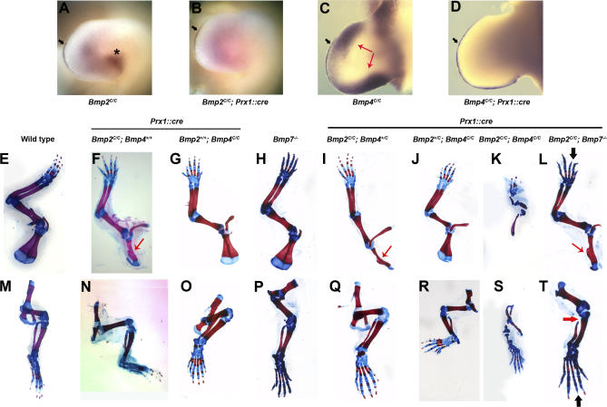
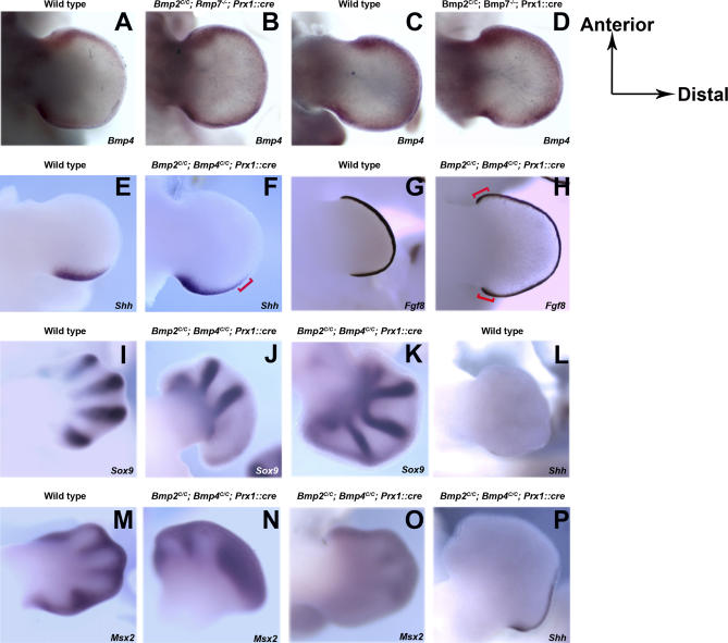
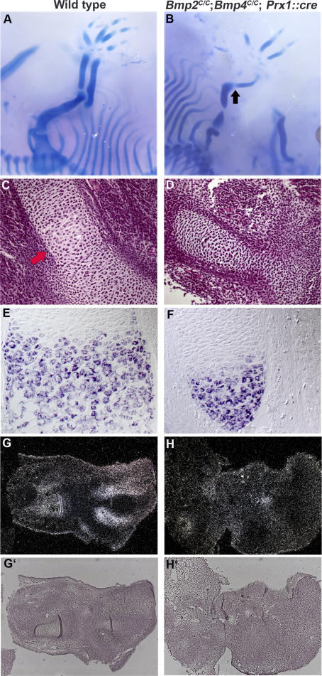
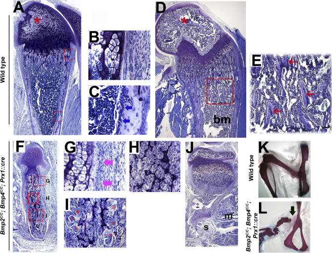
Bone morphogenetic protein (BMP) family members, including BMP2, BMP4, and BMP7, are expressed throughout limb development. BMPs have been implicated in early limb patterning as well as in the process of skeletogenesis. However, due to complications associated with early embryonic lethality, particularly for Bmp2 and Bmp4, and with functional redundancy among BMP molecules, it has been difficult to decipher the specific roles of these BMP molecules during different stages of limb development. To circumvent these issues, we have constructed a series of mouse strains lacking one or more of these BMPs, using conditional alleles in the case of Bmp2 and Bmp4 to remove them specifically from the limb bud mesenchyme. Contrary to earlier suggestions, our results indicate that BMPs neither act as secondary signals downstream of Sonic Hedghog (SHH) in patterning the anteroposterior axis nor as signals from the interdigital mesenchyme in specifying digit identity. We do find that a threshold level of BMP signaling is required for the onset of chondrogenesis, and hence some chondrogenic condensations fail to form in limbs deficient in both BMP2 and BMP4. However, in the condensations that do form, subsequent chondrogenic differentiation proceeds normally even in the absence of BMP2 and BMP7 or BMP2 and BMP4. In contrast, we find that the loss of both BMP2 and BMP4 results in a severe impairment of osteogenesis.


Conflict of interest statement

Competing interests. The authors have declared that no competing interests exist.

Figures

Figure 1
Figure 1. An Allelic/Nonallelic Series of BMP-Deficient Limbs
(A–D) Prx1::cre efficiently recombines Bmp2 and Bmp4 conditional alleles in the limbs. Bmp2 (A and B) and Bmp4 (C and D) whole mount mRNA in situ hybridization in the limb. Wild-type (A) and Bmp2C/C; Prx1::cre (B) are forelimbs from E10.5 mouse embryos. Mesenchymal expression [asterisk (A)] of Bmp2 is abolished in Bmp2C/C; Prx1::cre embryo while the AER expression [black arrow (A and B)] of Bmp2 persists. Note that pink staining in the central region of the limb bud in (B) is nonspecific background. Wild-type (C) and Bmp4C/C; Prx1::cre (D) are forelimbs from E10.5 mouse embryos. Mesenchymal expression [red arrow (C)] of Bmp4 is abolished in Bmp4C/C; Prx1::cre embryo while the AER expression [black arrow (C and D)] of Bmp4 persists. (E–T) Depletion of BMP2 and BMP4 together causes severe limb skeletal defects. (E–T) Whole mount skeletons from newborn animals stained with Alcian blue and Alizarin red. (E–L) Forelimbs, (M–T) hindlimbs. (E and M) Wild-type, (F and N) Bmp2C/C; Prx1::cre, (G and O) Bmp4C/C; Prx1::cre, (H and P) Bmp7/, (I and Q) Bmp2C/C; Bmp4+/C; Prx1::cre, (J and R) Bmp2+/C; Bmp4C/C; Prx1::cre, (K and S) Bmp2C/C; Bmp4C/C; Prx1::cre, (L and T) Bmp2C/C, Bmp7/; Prx1::cre. Thin red arrow in (F), (I), and (L), defective scapula; thick red arrow in (T), failure of fibula to articulate with knee, and thick black arrow in (L) and (T), missing phalanx in digit III.
Figure 2
Figure 2. Depletion of BMP Signaling Causes Interdigital Syndactyly
(A–D) Forelimb of adult wild-type (A) and Bmp2C/C; Prx1::cre mouse (B) and hindlimbs of newborn wild-type (C) mouse and newborn Bmp2C/C; Bmp4C/C; Prx1::cre (D) mouse. The black arrow in (B) shows soft tissue syndactyly in Bmp2C/C; Prx1::cre mouse. (E and F) Wild-type and Bmp2C/C; Bmp4C/C; Prx1::cre, respectively, showing acridine orange–stained hindlimbs of E15.5 mouse embryos. Acridine orange stain is in yellow. (G and H) Enlarged views of selected regions from (E) and (F), respectively. Black arrow in (G) and (H) show acridine orange–stained apoptotic cells in the interdigital mesenchyme, and asterisk in (H) shows the remnant of the AER. (I and J) Fgf8 mRNA expression in the hindlimbs of E13.5 wild-type (I) and Bmp2C/C; Bmp4C/C; Prx1::cre (J) embryos. The thick black arrows in (J) show Fgf8 mRNA expression.
Figure 3
Figure 3. Patterning Defects in Limbs Deficient of Different Combinations of BMP Molecules
(A–D) Bmp4 expression in limb buds from E11.5 wild-type (A and C) and Bmp2C/C, Bmp7/; Prx1::cre (B and D) mouse embryos. (A and B) Forelimbs, (C and D) hindlimbs. (E–H) Shh (E and F) and Fgf8 (G and H) expression in E11.5 forelimbs and hindlimbs, respectively. (E and G) Wild-type embryos, (F and H) Bmp2C/C; Bmp4C/C; Prx1::cre embryos. (I–P) Sox9 (I–K) and Msx2 (M–O) expression in E12.5 wild-type (I and M) and Bmp2C/C; Bmp4C/C; Prx1::cre mouse embryonic forelimbs (J and N) and hindlimbs (K and O). (L and P) Shh expression in E12.5 wild-type (L) and Bmp2C/C; Bmp4C/C; Prx1::cre (P) embryonic hindlimbs. The red brackets in (F) and (H) show the broadened domains of expressions of Shh and Fgf8, respectively.
Figure 4
Figure 4. Chondrogenesis Starts and Proceeds Normally Even in the Absence of BMP2 and BMP4
(A and B) Whole mount skeletons from E13.5 embryos that are stained with alcian blue. (A) Wild-type embryo, (B) Bmp2C/C; Bmp4C/C; Prx1::cre embryo. Black arrow in (B) shows the fusion of the zeugopod and stylopod. (C and D) Hematoxylin and eosin–stained sagittal sections of humeri from E13.5 wild-type (C) and Bmp2C/C; Bmp4C/C; Prx1::cre (D) embryos. Thick red arrow in (C) shows the hypertrophic region. (E and F) Sagittal sections of wild-type (E) and Bmp2C/C; Bmp4C/C; Prx1::cre (F) humeri from E15.5 embryos were hybridized with digoxigenin-labeled antisense rioprobes for ColX. (G and H) Sagittal sections of forelimbs from E13.5 wild-type and BMP2, BMP4–deficient embryos, respectively, stained with radioactive riboprobes for Bmp7 mRNA. (G′) and (H′) show the bright field views of (G) and (H), respectively.
Figure 5
Figure 5. In the Absence of BMP2 and BMP4, Osteogenesis Begins During Early Embryonic Development
(A–D) Sagittal sections of forelimb from E15.5 embryos. (A and B) Stained with toluidine blue. (A) Distal ulna from wild-type embryo, (B) distal ulna/radius from Bmp2C/C; Bmp4C/C; Prx1::cre embryo. Mineralized cartilage is shown by dark purple and osteoid is shown by light blue (red asterisk). (C and D) Humeri sections were hybridized with digoxigenin labeled riboprobe for ColI. (C) Section of wild-type humerus from E15.5 embryo, (D) from Bmp2C/C; Bmp4C/C; Prx1::cre E15.5 embryo. (E–H) Sagittal sections of zeugopod from E17.5 embryos. (E and F) Stained with toluidine blue while G and H are stained for TRAP (brown stain, red arrow). (E and G) E17.5 proximal radius from wild-type embryo, (F and H) E17.5 proximal ulna/radius from Bmp2C/C; Bmp4C/C; Prx1::cre embryo. Although vascularization occurs in the absence of BMP2 and BMP4, few osteoclasts are observed in the mineralized cartilage in Bmp2C/C; Bmp4C/C; Prx1::cre mice. Please note all the panels other than (G) and (H) are photographed at ×20, while (G) and (H) were photographed at ×40. (I and J) Sections of newborn proximal radius (I) from control animal and newborn proximal ulna/radius (J) from Bmp2C/C; Bmp4C/C; Prx1::cre animal were stained with toluidine blue. Red arrow shows the bone marrow cavity in (E) and (I). Note that bone collar (red *) forms at the right time and place in the absence of BMP2 and BMP4. Also note that since Bmp2C/C; Bmp4C/C; Prx1::cre mice occasionally have only one bone in zeugopod and the elbow joint is occasionally fused, it is difficult to distinguish between the ulna and the radius.
Figure 6
Figure 6. Defects in Bone Formation in the Absence of BMP2 and BMP4
Toluidine blue staining of sagittal sections of distal femurs. (A–E) Control femurs at 1 wk (A) and 3 wks (D) of age. Boxed areas in (A) are enlarged in (B) and (C). Boxed area in (D) is enlarged in (E). Blue arrows in (C) point to osteoblast cells lining the surface of cortical bone. Pink arrows in (G) show similar cells to those in (C) in Bmp2C/C; Bmp4C/C; Prx1::cre femurs. Red star in (A) and (D) marks the secondary ossification center. Pale blue–stained tissue in (E), marked by red arrows, is trabecular bone. bm, marks the bone marrow cavity in (D). (F–J) Bmp2C/C; Bmp4C/C; Prx1::cre femurs at 1 wk (F) and 3 wks (J) of age. Boxed areas in (F) are enlarged in (G), (H), and (I). (A), (D), (F), and (J) are shown with the same magnification. Note that there are defects in bone formation but no defect in osteoclast mediated bone resorption. Mineralized tissue in the mid shaft of femur is almost resorbed at 3 wks. (H) Fibroblast-like cells present in the bone shaft of Bmp2C/C; Bmp4C/C; Prx1::cre mouse. Red star in (I) shows the osteoclasts invading the Bmp2C/C; Bmp4C/C; Prx1::cre femurs at 1 wk of age. s and m mark soft tissues and muscle in (J), respectively. (K and L) Three-wk-old wild-type (K) and Bmp2C/C; Bmp4C/C; Prx1::cre (L) hindlimb skeletons stained with Alcian blue and Alizarin red. The black arrow shows the missing part of proximal femur.
Figure 7
Figure 7. Osteoblast Maturation Is Inhibited in the Absence of BMP2 and BMP4
(A–D) Distal femurs from control mice at 1 wk of age. (E–H) Distal femurs from Bmp2C/C; Bmp4C/C; Prx1::cre mice at 1 wk of age. (I–L) Distal femurs from control mice at 3 wks of age. (M–P) Distal femurs from Bmp2C/C; Bmp4C/C; Prx1::cre mice at 3 wks of age. In situ hybridization of early osteoblast differentiation-related marker genes Col I (B, F, J, and N), runx2 (C, G, K, and O) and osterix (D, H, L, and P).

References

    1. Reddi AH, Huggins C. Biochemical sequences in the transformation of normal fibroblasts in adolescent rats. Proc Natl Acad Sci U S A. 1972;69:1601–1605. - PMC - PubMed
    1. Urist MR. Bone: Formation by autoinduction. Science. 1965;150:893–899. - PubMed
    1. Wozney JM, Rosen V, Celeste AJ, Mitsock LM, Whitters MJ, et al. Novel regulators of bone formation: Molecular clones and activities. Science. 1988;242:1528–1534. - PubMed
    1. Hogan BL. Bone morphogenetic proteins: Multifunctional regulators of vertebrate development. Genes Dev. 1996;10:1580–1594. - PubMed
    1. Riddle RD, Johnson RL, Laufer E, Tabin C. Sonic hedgehog mediates the polarizing activity of the ZPA. Cell. 1993;75:1401–1416. - PubMed

Publication types

MeSH terms

LinkOut - more resources