. 2007 Jan 9;104(2):410-7.
doi: 10.1073/pnas.0610770104. Epub 2006 Dec 28.

Lung development and repair: contribution of the ciliated lineage

Affiliations

Lung development and repair: contribution of the ciliated lineage

Emma L Rawlins et al. Proc Natl Acad Sci U S A. .

Abstract

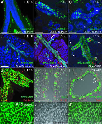
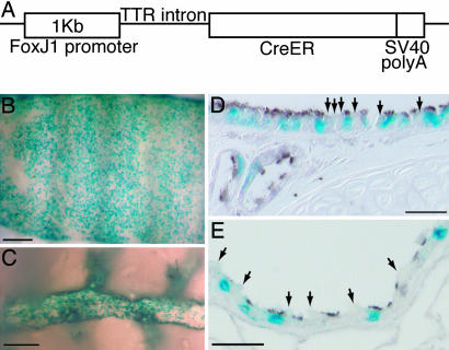
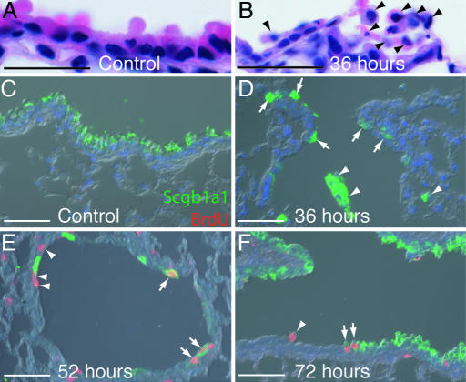
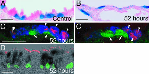
The identity of the endogenous epithelial cells in the adult lung that are responsible for normal turnover and repair after injury is still controversial. In part, this is due to a paucity of highly specific genetic lineage tools to follow efficiently the fate of the major epithelial cell populations: the basal, secretory, ciliated, neuroendocrine, and alveolar cells. As part of a program to address this problem we have used a 1-kb FOXJ1 promoter to drive CreER in the ciliated cells of the embryonic and adult lung. Analysis of FOXJ1-GFP transgenic lungs shows that labeled cells appear in a proximal-distal pattern during embryogenesis and that the promoter drives expression in all ciliated cells. Using FOXJ1CreER adult mice, we have followed the fate of ciliated cells after epithelial injury by naphthalene or sulfur dioxide. From quantitative analysis and confocal microscopy we conclude that ciliated cells transiently change their morphology in response to lung injury but do not proliferate or transdifferentiate as part of the repair process.


Conflict of interest statement

The authors declare no conflict of interest.

Figures

Fig. 1.
Fig. 1.
Ciliated cell patterning during development. (A–G) One hundred-micrometer vibratome sections of FOXJ1-GFP transgenic lungs. (A–E) Green, FOXJ1-GFP; red, anti-Scgb1a1; blue, anti-E-cadherin. (A) E13.5 mainstem-bronchi. FOXJ1-GFP is not yet expressed (green staining in the mesenchyme is nonspecific). (B) E14.5 mainstem-bronchus. FOXJ1-GFP is expressed in many cells. (C) E14.5 distal bronchiole from the same lung. FOXJ1-GFP-expressing cells are more scattered and are absent from distal tubules (∗). (D) E15.0 lung lobe. FOXJ1-GFP is expressed in scattered cells in the bronchioles but does not extend to the branching tips (brackets). (E) E15.5. FOXJ1-GFP is expressed in more cells but still does not extend to the branching tips (brackets). Scgb1a1 is first detectable at this stage by immunostaining. (F) E16.5. Green, FOXJ1-GFP; red, anti-Scgb1a1; blue, anti-CGRP. FOXJ1-GFP expression extends to the terminal bronchioles (arrows). Almost every epithelial cell expresses one of the three markers, but no cell expressing more than one marker was detected (the small amount of yellow labeling in the image is a result of boosting Scgb1a1 detection to visualize the more weakly expressing cells). Note that some CGRP-expressing cells are surrounded by clusters of Scgb1a1+ cells (∗). (G) E17.5. FOXJ1-GFP (green) colocalizes with anti-β-tubulin, a marker of surface cilia (red). (H) E15.0. Seven-micrometer paraffin section through an intralobular airway after 1 h of BrdU exposure. Green, anti-BrdU; red, anti-FoxJ1. The BrdU label does not colocalize with FoxJ1. (I) FOXJ1-GFP postnatal stage 3 weeks; 7-μm paraffin section through a bronchiole after 1 h of BrdU exposure. Green, anti-GFP; red, anti-BrdU. Proliferating cells (arrows) do not colocalize with FoxJ1. (J and J″) Whole-mount view of adult FOXJ1-GFP (green in J) bronchiole stained with tomato lectin (grayscale in J′) to visualize cell surfaces including cilia. The merged image (J″) shows that FOXJ1-GFP is expressed in all of the ciliated cells. (Scale bars: A–C, 100 μm; D–G, 200 μm; H, 20 μm; I and J, 40 μm.)
Fig. 2.
Fig. 2.
FOXJ1CreER2T transgene efficiently lineage-labels ciliated cells after tamoxifen injection. (A) FOXJ1CreER2T transgene construct. (B–E) X-gal-stained (blue) FOXJ1-CreER2T; Rosa26R adult airways after tamoxifen injection. (B) Whole-mount ventral trachea. (C) Intralobular airway. (D and E) Sections of anti-β-tubulin-stained trachea (brown) (D) and distal bronchiole (E). Most ciliated cells but no Clara cells (arrows) are lineage-labeled. (Scale bars: B and D, 200 μm; C and E, 20 μm.)
Fig. 3.
Fig. 3.
Characterization of the extent of the naphthalene injury and timing of proliferation. (A–F) Paraffin sections of bronchiole epithelium from male mice. (A and B) Hematoxylin and eosin staining. (A) Thirty-six hours after control corn oil injection. (B) Thirty-six hours after naphthalene. Arrowheads mark cells with no attachment to the basal lamina. (C–F) Anti-Scgb1a1 (green), anti-BrdU (red), and DAPI (blue). (C) Fifty-two hours after control injection. Clara cells predominate the airway epithelium and no dividing cells are present. (D) Thirty-six hours after naphthalene. Clara cells have detached from the basal lamina (arrowheads), but small numbers are retained at the BADJ (arrows) and at intervals throughout the bronchioles (data not shown). Cells have not yet started to divide. (E) Fifty-two hours after naphthalene injection. Dividing epithelial cells are observed; they are either Scgb1a1+ (arrows) or Scgb1a1 (arrowheads). (F) Seventy-two hours after naphthalene injection. Dividing Scgb1a1+ (arrows) and Scgb1a1 (arrowhead) cells are still observed, and many more Clara cells are also present. (Scale bars: A and B, 20 μm; C–F, 200 μm.)
Fig. 4.
Fig. 4.
Response of ciliated cells to naphthalene injury. (A–D) FOXJ1-CreER2T; Rosa26R 7-μm paraffin sections. (A and B) X-gal-stained (blue) and eosin-stained (pink) bronchioles. (A) Fifty-two hours after control corn oil injection. (B) Fifty-two hours after naphthalene. Domed Clara cells are absent, and X-gal+ cells are flattened. (C and C′) Fifty-two hours after naphthalene. Two confocal z-sections of the same BADJ. Green, anti-β-gal; red, anti-BrdU; blue, anti-Scgb1a1. Lineage-labeled cells (arrows) have not incorporated BrdU. Proliferating cells are Scgb1a1+ (arrowheads). (D) Proximal tracheal section 52 h after naphthalene injury. Black cytoplasm, X-gal; red cilia, anti-β-tubulin; green nuclei, anti-BrdU. Lineage-labeled cells have not incorporated BrdU. (Scale bars: 20 μm.)
Fig. 5.
Fig. 5.
Ciliated cells do not transdifferentiate after naphthalene injury. FOXJ1-CreER2T; Rosa26R airways. (A) One hundred days after naphthalene exposure. Whole-mount image of X-gal-stained (blue), microdissected bronchiole. The X-gal+ cells are tightly clustered and separated by regions of unlabeled cells. Compare with Fig. 2C. (B–E) Seven-micrometer paraffin sections of bronchioles 3 weeks after naphthalene injury. (B) X-gal (blue) and anti-β-tubulin (brown). Ninety-eight percent of lineage-labeled cells are clearly β-tubulin+ (arrows). Domed Clara cells are not lineage-labeled (arrowheads). Two percent of lineage-labeled cells do not have visible cilia (red arrowhead). (C–E) Single confocal z-sections of lineage-labeled X-gal+ cells (black) and Scgb1a1+ cells (red). (C and D) Typical bronchioles. Lineage-labeled cells are Scgb1a1 (arrows). (E) One percent of lineage-labeled cells colabel with Scgb1a1 (arrowheads). (Scale bars: A, 200 μm; B, 40 μm; C, 60 μm; D and E, 20 μm.)
Fig. 6.
Fig. 6.
Characterization of the extent of SO2 injury and timing of proliferation. (A–C) Transverse tracheal sections. (A) Uninjured control. (B) Six hours after SO2 inhalation. Epithelial cells are sloughing off. (C) Twenty-four hours after SO2 inhalation. The luminal epithelium consists of a monolayer of low cuboidal cells. (D) Percentage of epithelial cells (in the most proximal one-third of the trachea) that incorporated BrdU in a 1-h pulse at different times after the injury. Bars are the standard error of the mean. Epithelial cell proliferation peaks 24 h after the injury. (Scale bars: 200 μm.)
Fig. 7.
Fig. 7.
Ciliated cells after SO2 inhalation. Shown are FOXJ1-CreER2T; Rosa26R ventral trachea. Blue, X-gal. (A–E) Whole-mount images of ventral tracheal epithelial surface. (F–I) Seven-micrometer paraffin sections taken from the regions between the dotted lines. (A and F) Tamoxifen-treated positive control. X-gal+ cells (arrows) are in the luminal epithelium and SMG ducts (∗). (B and G) Negative control (no tamoxifen injections). Low level of endogenous X-gal staining in the SMG mucus cells (arrowhead). (C and H) Tamoxifen-treated, 24 h after SO2 inhalation. X-gal+ cells (arrows) are restricted to the SMG ducts. (D and I) Tamoxifen-treated, 2 weeks after SO2 inhalation. X-gal+ cells (arrows) are still restricted to the SMG ducts. (E) Tamoxifen-treated, 24 h after SO2 inhalation. In some animals, the surviving X-gal+ cells are scattered throughout the luminal epithelium, particularly in the distal region. (Scale bars: A–E, 0.5 mm; G–I, 10 μm.)
Fig. 8.
Fig. 8.
Ciliated cells do not divide or transdifferentiate after SO2 exposure. Shown are FOXJ1-CreER2T; Rosa26R X-gal-stained 7-μm paraffin sections of the trachea. (A and B) Twenty-four hours after SO2 inhalation. (A) Proximal trachea with SMG duct. Anti-BrdU (black) and X-gal (blue; arrows) do not colocalize. (B) Single confocal z-section of distal trachea. Anti-BrdU (red) and X-gal (black) do not colocalize. (C and D) Two weeks after SO2 inhalation. Black, anti-β-tubulin; blue, X-gal. (C) Typical trachea section; ciliated cells have differentiated in the repaired epithelium but are not lineage-labeled. (D) Lineage-labeled cells have cilia (arrows). Domed Clara cells have not inherited the lineage label (arrowheads). (Scale bars: 20 μm.)

References

    1. Rawlins EL, Hogan BL. Development (Cambridge, UK) 2006;133:2455–2465. - PubMed
    1. Neuringer IP, Randell SH. Monaldi Arch Chest Dis. 2006;65:47–51. - PubMed
    1. Cardoso WV, Lu J. Development (Cambridge, UK) 2006;133:1611–1624. - PubMed
    1. Okubo T, Knoepfler PS, Eisenman RN, Hogan BL. Development (Cambridge, UK) 2005;132:1363–1374. - PubMed
    1. Perl AK, Wert SE, Nagy A, Lobe CG, Whitsett JA. Proc Natl Acad Sci USA. 2002;99:10482–10487. - PMC - PubMed

Publication types

MeSH terms

LinkOut - more resources