. 2007 Jan 9;104(2):456-61.
doi: 10.1073/pnas.0607817104. Epub 2006 Dec 28.

Structure of aspartoacylase, the brain enzyme impaired in Canavan disease

Affiliations

Structure of aspartoacylase, the brain enzyme impaired in Canavan disease

Eduard Bitto et al. Proc Natl Acad Sci U S A. .

Abstract

Aspartoacylase catalyzes hydrolysis of N-acetyl-l-aspartate to aspartate and acetate in the vertebrate brain. Deficiency in this activity leads to spongiform degeneration of the white matter of the brain and is the established cause of Canavan disease, a fatal progressive leukodystrophy affecting young children. We present crystal structures of recombinant human and rat aspartoacylase refined to 2.8- and 1.8-A resolution, respectively. The structures revealed that the N-terminal domain of aspartoacylase adopts a protein fold similar to that of zinc-dependent hydrolases related to carboxypeptidases A. The catalytic site of aspartoacylase shows close structural similarity to those of carboxypeptidases despite only 10-13% sequence identity between these proteins. About 100 C-terminal residues of aspartoacylase form a globular domain with a two-stranded beta-sheet linker that wraps around the N-terminal domain. The long channel leading to the active site is formed by the interface of the N- and C-terminal domains. The C-terminal domain is positioned in a way that prevents productive binding of polypeptides in the active site. The structures revealed that residues 158-164 may undergo a conformational change that results in opening and partial closing of the channel entrance. We hypothesize that the catalytic mechanism of aspartoacylase is closely analogous to that of carboxypeptidases. We identify residues involved in zinc coordination, and propose which residues may be involved in substrate binding and catalysis. The structures also provide a structural framework necessary for understanding the deleterious effects of many missense mutations of human aspartoacylase.


Conflict of interest statement

The authors declare no conflict of interest.

Figures
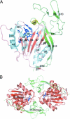
Fig. 1.
Fig. 1.
Ribbon diagrams of the rASPA monomer and dimer. (A) N-domain of rASPA is color-coded in cyan and red. C-domain is color-coded in yellow and green. Residues His-21, Gly-22, Glu-24, Asn-54, Arg-63, Asn-70, Arg-71, Phe-73, Asp-114, His-116, and Glu-178 (blue sticks) are highly conserved in the AstE-AspA family and delineate the active site. Zn2+ is shown as a pink sphere. (B) The rASPA dimer observed in the asymmetric unit of the rASPA crystals is shown in ribbon representation. Both the N-domain (red) and C-domain (green) of the rASPA monomers are involved in formation of the dimer interface. Residues His-21, Glu-24, and His-116 (blue sticks) coordinate Zn2+ (pink sphere).
Fig. 2.
Fig. 2.
Details of the ASPA active site. (A) A stereo representation of structural superposition of rASPA (red) and bovine carboxypeptidase A (cyan; PDB code 1m4l). (B) A stereo representation of structural superposition of the active sites of bovine pancreatic carboxypeptidase A (cyan, PDB code 6cpa) and rASPA (red). Red and cyan lines represent the Cα-trace of the enzymes. Important residues involved in Zn2+ (sphere) coordination, substrate binding, and catalysis are shown in sticks and are annotated for hASPA (corresponding carboxypeptidase A residues are listed in Table 2). A transition state inhibitor (blue lines) is bound in the active site of carboxypeptidase A. Selected atoms of this inhibitor that topologically correspond to those of putative NAA transition state are highlighted (magenta) for clarity. Sulfate bound in the active site of rASPA is show in orange sticks. Arg-71 of rASPA adopts two alternate conformations. (C) The 2FoFc electron density map (blue) of rASPA is shown at contour level of 1.8σ. rASPA residues are shown in sticks, the metal ion (magenta) coordinated in the active site and waters (red) are depicted by three-dimensional crosses. (D) The 2FoFc electron density map (blue) of hASPA is shown at contour level of 1.5σ. The anomalous difference map (red) is shown at contour level of 6σ. Black dashed lines represent coordination bonds from protein residues and phosphate to Zn2+ (magenta) bound in the active site of hASPA.
Fig. 3.
Fig. 3.
Comparison of the substrate binding cavities. Lines represent Cα-traces of the rASPA (red) and carboxypeptidase A (cyan). Both proteins were superposed and are shown in the same orientation. Solvent accessible electrostatic surfaces of both proteins are clipped to reveal internal pockets responsible for binding of substrates (black sticks, see Fig. 2B for details). Arrows indicate direction in which substrates enter binding pockets of both proteins. Zinc ions are shown as spheres. (A) The substrate binding cavity is formed at the interface of the N- and C-domains of rASPA. (B) The substrate binding cavity recognizes the terminal hydrophobic and a portion of the penultimate residue of the substrate in bovine pancreatic carboxypeptidase A (PDB ID code 6cpa).
Fig. 4.
Fig. 4.
Proposed mechanism of action of ASPA.

Comment in

  • The impact of structural biology on neurobiology.
    Viola RE. Viola RE. Proc Natl Acad Sci U S A. 2007 Jan 9;104(2):399-400. doi: 10.1073/pnas.0610164103. Epub 2007 Jan 3. Proc Natl Acad Sci U S A. 2007. PMID: 17213329 Free PMC article. No abstract available.

Similar articles

Cited by

References

    1. Baslow MH. Neurochem Res. 2003;28:941–953. - PubMed
    1. Clark JF, Doepke A, Filosa JA, Wardle RL, Lu A, Meeker TJ, Pyne-Geithman GJ. Med Hypotheses. 2006;67:506–512. - PubMed
    1. Tsai G, Coyle JT. Prog Neurobiol. 1995;46:531–540. - PubMed
    1. Gujar SK, Maheshwari S, Bjorkman-Burtscher I, Sundgren PC. J Neuroophthalmol. 2005;25:217–226. - PubMed
    1. Madhavarao CN, Chinopoulos C, Chandrasekaran K, Namboodiri MA. J Neurochem. 2003;86:824–835. - PubMed

Publication types

MeSH terms