. 2006 Dec;18(12):3721-44.
doi: 10.1105/tpc.106.044180. Epub 2006 Dec 28.

Phytotoxicity and innate immune responses induced by Nep1-like proteins

Affiliations

Phytotoxicity and innate immune responses induced by Nep1-like proteins

Dinah Qutob et al. Plant Cell. 2006 Dec.

Abstract

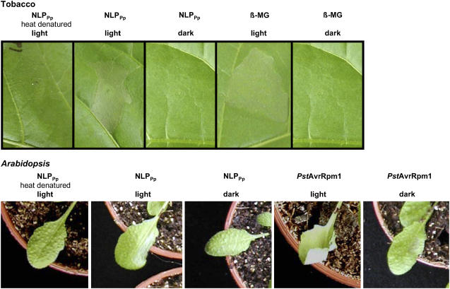
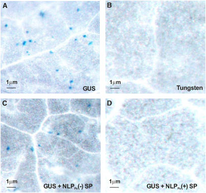
We show that oomycete-derived Nep1 (for necrosis and ethylene-inducing peptide1)-like proteins (NLPs) trigger a comprehensive immune response in Arabidopsis thaliana, comprising posttranslational activation of mitogen-activated protein kinase activity, deposition of callose, production of nitric oxide, reactive oxygen intermediates, ethylene, and the phytoalexin camalexin, as well as cell death. Transcript profiling experiments revealed that NLPs trigger extensive reprogramming of the Arabidopsis transcriptome closely resembling that evoked by bacteria-derived flagellin. NLP-induced cell death is an active, light-dependent process requiring HSP90 but not caspase activity, salicylic acid, jasmonic acid, ethylene, or functional SGT1a/SGT1b. Studies on animal, yeast, moss, and plant cells revealed that sensitivity to NLPs is not a general characteristic of phospholipid bilayer systems but appears to be restricted to dicot plants. NLP-induced cell death does not require an intact plant cell wall, and ectopic expression of NLP in dicot plants resulted in cell death only when the protein was delivered to the apoplast. Our findings strongly suggest that NLP-induced necrosis requires interaction with a target site that is unique to the extracytoplasmic side of dicot plant plasma membranes. We propose that NLPs play dual roles in plant pathogen interactions as toxin-like virulence factors and as triggers of plant innate immune responses.


Figures

Figure 1.
Figure 1.
Phylogeny of NLPs. The Nep1 protein sequence and 43 related sequences are shown. The scale bar represents 20% weighted sequence divergence. GenBank identifier numbers for each protein sequence are shown along with the species of origin. Sequences with special relevance to this study are additionally labeled: Nep1, necrosis and ethylene inducing peptide 1; NLPPp, NLP from Phytophthora parasitica; NLPPs, NLP from Phytophthora sojae; NLPPya, NLP from Pythium aphanidermatum.
Figure 2.
Figure 2.
NLP-Induced Activation of Plant Immune Responses in Arabidopsis. (A) Five-week-old Arabidopsis plants were infiltrated with 2 μM recombinant NLPPp or glutathione S-transferase (GST) as control (Fellbrich et al., 2002) for the times indicated. Proteins were extracted and subjected to protein blot analysis using a 1:1000 dilution of phospho-p44/42 MAP kinase antibody as described in Methods. (B) Arabidopsis cell culture aliquots (2.5 × 10⁴/50 μL) were treated with the indicated recombinant NLPPp concentrations or Escherichia coli protein extracts as control. NO production is given as relative fluorescence units (rfu). (C) Camalexin accumulation in 5-week-old Arabidopsis rosette leaves after infiltration with 2 μM recombinant NLPPs (black bars), 2 μM recombinant NLPPya (gray bars), or protein renaturation buffer as control (white bars). Camalexin was extracted at the time points indicated and determined as described in Methods. All experiments shown in (A) to (C) were performed at least three times with identical results. Data in (B) and (C) show average values + sd. (D) and (E) Transcriptome analysis in 5-week-old Arabidopsis plants treated with 1 μM recombinant NLPPp (GST as control) or 1 μM synthetic flg22 (water as control). All experiments were performed in triplicate, and expression levels for each probe set were analyzed as described in Materials. (D) Behavior of 12,557 genes significantly expressed at 1 or 4 h after treatments. Scatterplot analysis of fold induction of the probe sets for NLPPp trials (4 h) versus fold induction for flg22 trials (4 h). For each treatment versus control condition, genes that changed were assigned based on a one-way analysis of variance (ANOVA) test combined with a Benjamini and Hochberg false discovery rate algorithm. The numbers given in the inset refer to those genes of which expression was statistically significantly altered more than twofold at least at one of the two time points tested. The rectangular box comprises those genes that are unaltered in expression upon stimulation. Probe sets in areas enclosed by dotted lines are coordinately upregulated (red dots) or downregulated (green dots). (E) Venn diagram showing the total number of genes that are coordinately expressed by both stimuli (overlap) or of which expression is specifically upregulated by either NLPPp or flg22 treatment.
Figure 3.
Figure 3.
Germination of Arabidopsis Seedlings in the Presence of NLPPs. (A) Seeds of Arabidopsis ecotype Col-0 were sown in sterile media containing a range of concentrations of NLPPs, and root lengths were measured at intervals as indicated. Shown are average values and sd from measurements of 10 to 20 plants (per treatment) from a representative experiment. The experiment was performed three times with similar results. (B) Seeds of 17 Arabidopsis ecotypes were sown in sterile media, with and without added NLPPs, and root lengths were measured after 8 d. Shown are average values and sd from measurements of 10 to 20 plants (per ecotype) from a representative experiment. The experiment was performed three times with similar results. (C) Seeds of Arabidopsis ecotype Col-0 were sown in sterile half-strength MS medium alone (top panel), on half-strength MS medium supplemented with 1.0 μg/mL NLPPs (middle panel), or on half-strength MS medium containing 1.0 μg/mL heat-denatured NLPPs (bottom panel). Photographs were taken 5 d after sowing.
Figure 4.
Figure 4.
NLP-Induced Cell Death Requires Active Plant Metabolism. Four-week-old tobacco plants were infiltrated either with the calcium channel blocker LaCl3 (1 mM), with the DNA transcription inhibitor α-amanitin (100 μM), or the protein translation inhibitor cycloheximide (CHX; 100 μM) alone, in combination with buffer as control, or 1 μM NLPPp or 50 nM β-megaspermin (β-MG), respectively. PCD symptoms shown here were obtained after 2 d.
Figure 5.
Figure 5.
Light Dependence of NLPPp-Induced Cell Death. Five-week-old tobacco (top panel) or Arabidopsis plants (bottom panel) were treated with 1 μM NLPPp, 1 μM heat-denatured NLPPp, 50 nM β-megaspermin (β-MG), or 5 × 10⁶ colony-forming units/mL P. syringae pv tomato strain DC3000/AvrRpm1 (PstAvrRpm1) under normal light conditions or 30 min upon transfer into the dark as indicated. PCD symptoms shown here were obtained after 2 d.
Figure 6.
Figure 6.
NLP-Induced PCD Is Independent of Caspase and Bax Inhibitor Activity. (A) Tobacco leaves were infiltrated with 4 μM NLPPp or P. syringae pv phaseolicola strains NPS4000 (PCD-noninducing strain) or NSP3121 (PCD-inducing strain) in the presence or absence of 100 μM of the caspase inhibitors Ac-YVAD-CHO, Ac-DEVD-CHO, or zVAD-FMK. Plants were kept at room temperature under continuous illumination. (B) Five-week-old Arabidopsis Col-0, atbi1-1, or atbi1-2 mutant plants were treated with 4 μM NLPPp, 4 μM heat-denatured NLPPp, or buffer as control. Photographs in (A) and (B) were taken 24 h after infiltration.
Figure 7.
Figure 7.
NLP-Induced Cell Death in Dicot Plants Does Not Require Intact Plant Cell Walls. Parsley, Arabidopsis, or maize protoplasts (5 × 10⁵/mL) prepared from cultured cells were treated with 1 μM NLPPp for 24 h, and viability staining was performed as described in Methods.
Figure 8.
Figure 8.
NLPs Possess Binding Affinity to Phospholipid Bilayers but Do Not Show Pore-Forming Activity on Artificial and Biological Membranes. (A) TRANSIL beads coated with POPE/POPC (20:80), POPE/POPC (5:95), or POPC alone were incubated for 1 h with 1 μM NLPPp (total protein [T]). After separation of lipid-bound (B) from free unbound material (F), proteins were analyzed by SDS-PAGE and silver staining. (B) Na+ influx into Sodium Green–filled liposomes. Sodium-mediated fluorescence was measured without protein and in the presence of 1 μM NLPPp (open symbols) or 1 μM HrpZPsph (closed symbols). Fluorescence values obtained without protein were subtracted from values obtained in the presence of either protein. Values given represent the means ± sd from assays performed in triplicate. (C) Two-electrode voltage clamp measurements on X. laevis oocytes. The current/voltage plots obtained before and after the application of 1 μM NLPPya (open squares and closed circles, respectively) or 1 μM HrpZPsph (open circles) are shown. Steady state currents were measured following 4-s pulses. The results presented are representative of those obtained in three experiments ±sd.
Figure 9.
Figure 9.
NLP-Induced PCD Requires Delivery to and Recognition at the Extracytoplasmic Side of Dicotyledonous Plant Cells. Activity of NLPPs in Arabidopsis leaves as determined by a cobombardment and transient expression assay. The photographs show Arabidopsis leaves after bombardment with tungsten beads and histochemical staining for GUS activity, performed as described in Methods. The tungsten beads were treated as follows: (A), coated with pFF19G containing a GUS expression cassette; (B), uncoated tungsten beads alone; (C), coated with pFF19G (GUS) plus a pFF19 construct encoding NLPPs lacking a signal peptide [NLPPs(-)SP]; (D), coated with pFF19G (GUS) plus a pFF19 construct encoding NLPPs, including the complete open reading frame with the native signal peptide [NLPPs(+)SP].

Similar articles

Cited by

References

    1. Akira, S., Uematsu, S., and Takeuchi, O. (2006). Pathogen recognition and innate immunity. Cell 124 783–801. - PubMed
    1. Alfano, J.R., and Collmer, A. (2004). Type III secretion system effector proteins: Double agents in bacterial disease and plant defense. Annu. Rev. Phytopathol. 42 385–414. - PubMed
    1. Amsellem, Z., Cohen, B.A., and Gressel, J. (2002). Engineering hypervirulence in a mycoherbicidal fungus for efficient weed control. Nat. Biotechnol. 20 1035–1039. - PubMed
    1. Asai, T., Stone, J.M., Heard, J.E., Kovtun, Y., Yorgey, P., Sheen, J., and Ausubel, F.M. (2000). Fumonisin B1-induced cell death in Arabidopsis protoplasts requires jasmonate-, ethylene-, and salicylate-dependent signaling pathways. Plant Cell 12 1823–1836. - PMC - PubMed
    1. Asai, T., Tena, G., Plotnikova, J., Willmann, M.R., Chiu, W.L., Gomez-Gomez, L., Boller, T., Ausubel, F.M., and Sheen, J. (2002). MAP kinase signalling cascade in Arabidopsis innate immunity. Nature 415 977–983. - PubMed

Publication types

MeSH terms