. 2006 Dec;18(12):3576-93.
doi: 10.1105/tpc.105.040774. Epub 2006 Dec 28.

The essential nature of sphingolipids in plants as revealed by the functional identification and characterization of the Arabidopsis LCB1 subunit of serine palmitoyltransferase

Affiliations

The essential nature of sphingolipids in plants as revealed by the functional identification and characterization of the Arabidopsis LCB1 subunit of serine palmitoyltransferase

Ming Chen et al. Plant Cell. 2006 Dec.

Abstract

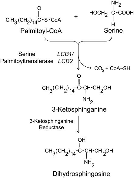
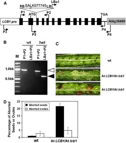
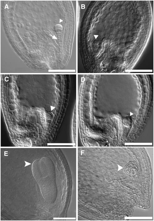
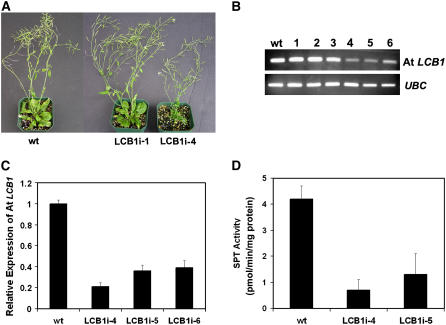
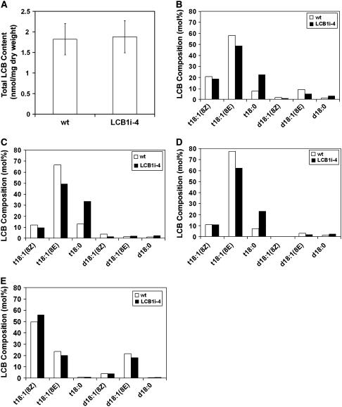
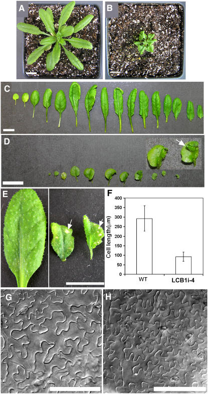
Serine palmitoyltransferase (SPT) catalyzes the first step of sphingolipid biosynthesis. In yeast and mammalian cells, SPT is a heterodimer that consists of LCB1 and LCB2 subunits, which together form the active site of this enzyme. We show that the predicted gene for Arabidopsis thaliana LCB1 encodes a genuine subunit of SPT that rescues the sphingolipid long-chain base auxotrophy of Saccharomyces cerevisiae SPT mutants when coexpressed with Arabidopsis LCB2. In addition, homozygous T-DNA insertion mutants for At LCB1 were not recoverable, but viability was restored by complementation with the wild-type At LCB1 gene. Furthermore, partial RNA interference (RNAi) suppression of At LCB1 expression was accompanied by a marked reduction in plant size that resulted primarily from reduced cell expansion. Sphingolipid content on a weight basis was not changed significantly in the RNAi suppression plants, suggesting that plants compensate for the downregulation of sphingolipid synthesis by reduced growth. At LCB1 RNAi suppression plants also displayed altered leaf morphology and increases in relative amounts of saturated sphingolipid long-chain bases. These results demonstrate that plant SPT is a heteromeric enzyme and that sphingolipids are essential components of plant cells and contribute to growth and development.


Figures

Figure 1.
Figure 1.
Biosynthesis of Sphingolipid Long-Chain Bases. The first step in the synthesis of sphingolipid long-chain bases is the condensation of Ser and palmitoyl-CoA, which is catalyzed by SPT. Eukaryotic forms of SPT are composed of subunits designated LCB1 and LCB2. The 3-ketosphinganine product of SPT is reduced by 3-ketosphinganine reductase to form dihydrosphingosine (d18:0), the simplest long-chain base. Other long-chain bases are formed by further hydroxylation and desaturation of dihydrosphingosine.
Figure 2.
Figure 2.
At LCB1 Amino Acid Sequence Phylogeny and Properties. (A) Phylogenetic analysis of At LCB1 relative to LCB1, LCB2, and other closely related members of the α-oxoamine synthase subfamily of pyridoxal 5′-phosphate–dependent enzymes. Bootstrap values shown at nodes were obtained from 5000 trials, and branch lengths correspond to the divergence of sequences, as indicated by the relative scale. AONS, 8-amino-7-oxononanoate synthase; LCB1, SPT LCB1 subunit; LCB2, SPT LCB2 subunit; sSPT, soluble SPT. Species are as follows: At, Arabidopsis thaliana; Bc, Bacillus cereus; Ca, Candida albicans; Cg, Cricetulus griseus; Hs, Homo sapiens; Kl, Kluyveromyces lactis; Mm, Mus musculus; Os, Oryza sativa; Sc, Saccharomyces cerevisiae; St, Solanum tuberosum; Sp, Sphingomonas paucimobilis; Td, Treponema denticola; and Zm, Zymomonas mobilis. (B) Comparison of the pyridoxal phosphate binding motif of LCB1 and LCB2 of Arabidopsis and S. cerevisiae SPT. The Lys (arrow) that binds pyridoxal 5′-phosphate through a Schiff's base is absent from LCB1 subunits but is present in LCB2 and other α-oxoamine synthase–related polypeptides.
Figure 3.
Figure 3.
Coexpression of At LCB1 and At LCB2 Complements the Long-Chain Base Auxotrophy of S. cerevisiae LCB1 and LCB2 Single and Double Mutants. (A) The At LCB1 and At LCB2 genes were expressed in wild-type or lcb1Δ, lcb2Δ, and lcb1Δ lcb2Δ mutant S. cerevisiae cells. Shown are immunoblots of microsomes isolated from yeast cells. The yeast LCB1 (Sc LCB1) and At LCB1 proteins were detected using a polyclonal antibody against the yeast LCB1. The yeast LCB2 (Sc LCB2) and At LCB2 polypeptides were detected using polyclonal antibodies prepared against peptides from the corresponding proteins. (B) Growth of wild-type yeast or lcb1Δ, lcb2Δ, lcb1Δ lcb2Δ, and lcb1Δ tsc3Δ mutants expressing the At LCB1 and At LCB2 genes individually or together on galactose-containing medium without added phytosphingosine. pESC corresponds to cells harboring the empty expression vector. (C) SPT activity in microsomes from yeast lcb1Δ cells expressing At LCB1 and At LCB2 individually or together (n = 3; average ± sd). For comparison, SPT activity in microsomes from wild type yeast was 100 ± 10 pmol·min−1·mg−1 protein.
Figure 4.
Figure 4.
Expression of At LCB1 in Arabidopsis and Its Subcellular Localization. (A) RT-PCR analysis of At LCB1 expression in young leaves (YL), mature leaves (ML), stems (ST), flowers (F), siliques (Si), and roots (R). A ubiquitin-conjugating enzyme gene (UBC; At5g25760) was used as an internal control. (B) to (F) Localization of At LCB1 promoter–GUS activity in Arabidopsis transgenic plants. (B) Ten-day-old seedling. (C) High expression of GUS in guard cells of cotyledons (arrowheads). Bar = 100 μm. (D) Anthers. (E) Siliques at 2, 4, and 7 d after flowering. (F) GUS expression in central replum (white arrows) and funiculus (black arrowheads) from siliques at 7 d after flowering. (G) to (J) Subcellular localization of At LCB1 as revealed by transient expression in tobacco leaves. (G) Distribution of At LCB1-EYFP. (H) Distribution of the ER marker CSP-CFP-HDEL. (I) Merge of (G) and (H) showing colocalization of At LCB1-EYFP with the ER marker. (J) White light image of tobacco epidermal cells. Bar = 10 μm.
Figure 5.
Figure 5.
Gene Structure of At LCB1 and Characterization of the At lcb1-1 Mutant Allele. (A) Scheme of At LCB1. The predicted At LCB1 open reading frame contains 13 exons (black boxes) and 12 introns (black lines). The At LCB1 promoter (LCB1 pro) and neighboring gene (At4g36490) are also shown. The T-DNA insert in SALK_077745 (indicated by the inverted triangle) is located in intron 2 and has the same orientation to the At LCB1 gene. The primers shown were used to determine the location of the T-DNA insertion, to amplify the genomic sequence for complementation experiments, and to verify the complementation of the SALK_077745 mutant. (B) PCR genotyping of SALK_077745 T-DNA lines. PCR was conducted with genomic DNA from a population of SALK_077745 T4 plants using a pair of At LCB1–specific primers (P1 and P2) or by the combination of T-DNA left border–specific primers (LBa1) and a corresponding At LCB1–specific primer (P2). In wild-type plants, only the wild-type allele (indicated by an ∼900-bp band; white arrowhead) was amplified, and no T-DNA allele was detected. However, in the heterozygous At LCB1-1 (het) plants, both the wild-type allele (∼900 bp) and the T-DNA disruption allele (indicated by an ∼600-bp band; black arrowhead) were amplified. (C) Morphology of seeds from wild-type (Col-0) and heterozygous At lcb1-1 plants. Shown are siliques from a wild-type plant (top panel) and from heterozygous At lcb1-1 plants at 7 to 10 d after flowering (middle panel) and 14 d after flowering (bottom panel). The aborted seeds are either pale (arrowheads) or brown and shrunken (asterisks), depending upon the maturity of seeds. (D) Average percentage of aborted seeds and ovules from siliques of wild-type (Col-0) and selfed heterozygous At LCB1/At lcb1 plants. The average number of aborted seeds and ovules from 10 siliques collected from five plants and the sd are presented (n = 5; >500 total seeds were examined). Ovule abortion was assessed as described previously (Meinke, 1994).
Figure 6.
Figure 6.
Defective Embryo Development is Observed in Homozygous At lcb1-1 Seeds. (A) Wild-type seed (2 d after flowering) showing a globular embryo (arrowhead) and an extended suspensor (arrow). (B) to (D) Aberrant embryos observed in homozygous At lcb1-1 seeds. The homozygous segregants were dissected from siliques collected at 2 d after flowering from selfed heterozygous At lcb1-1 plants. (B) Embryo arrested at the one-cell stage. (C) Embryo arrested at the two-cell stage. (D) Early globular embryo with an abnormal cell pattern and a shortened suspensor. (E) Wild-type embryo at the torpedo stage (4 to 5 d after flowering). (F) Residue of a degenerated embryo from a homozygous At lcb1-1 seed (4 to 5 d after flowering). Bars = 100 μm.
Figure 7.
Figure 7.
RNAi-Mediated Suppression of At LCB1 Results in Distinct Growth Phenotypes That Correlate with the Relative Degree of Reduced At LCB1 Expression. (A) Two phenotypic classes were observed in T1 At LCB1 RNAi plants: one class that was indistinguishable from wild-type plants, as represented by LCB1i-1, and a second class with reduced overall size and curled leaves, as represented by LCB1i-4. (B) RT-PCR analyses of wild-type and representative At LCB1 RNAi suppression lines. Plants indistinguishable from wild-type plants had no detectable reduction in At LCB1 expression (lanes 1 to 3), whereas small plants with altered leaf morphology displayed reduced expression of At LCB1 (lanes 4 to 6). A gene for a ubiquitin-conjugating enzyme (see Methods) was used as an internal control. (C) Real-time PCR quantification of At LCB1 expression T2 plants from At LCB1 suppression lines. Expression of At LCB1 was normalized relative to the expression of a ubiquitin-conjugating enzyme gene. Results from three independent experiments are presented as averages ± sd. (D) SPT activity in microsomes from leaves of wild-type (Col-0) and LCB1i-4 and LCB1i-5 RNAi plants (n = 3; average ± sd).
Figure 8.
Figure 8.
Effects of At LCB1 RNAi Suppression on Sphingolipid Long-Chain Base Content and Composition. (A) Total sphingolipid long-chain base content of leaves from wild-type and LCB1i-4 RNAi suppression plants (n = 10; average ± sd). (B) Long-chain base composition of sphingolipids in the total solvent extract from leaves of wild-type and LCB1i-4 plants. Data in (B) to (E) are representative of three independent experiments. (C) Long-chain base composition of sphingolipids in the tissue residue after solvent extraction from leaves of wild-type and LCB1i-4 plants. (D) Long-chain base composition of charged sphingolipids from leaves of wild type and LCB1i-4 plants. This fraction is composed primarily of glycosylated inositolphosphoceramides (Markham et al., 2006). (E) Long-chain base composition of monoglucosylceramides from leaves of wild-type and LCB1i-4 plants.
Figure 9.
Figure 9.
RNAi-Mediated Suppression of At LCB1 Expression Results in Reduced Sizes of Plants and Cells and Altered Leaf Morphology. The data shown are from the LCB1i-4 line. Similar results were obtained with pHannibal-based At LCB1 RNAi lines. (A) and (B) Four-week-old wild-type (A) and LCB1i-4 RNAi suppression (B) plants. (C) Arrangement of all leaves from a 4-week-old wild-type plant. (D) Arrangement of all leaves from a 4-week-old LCB1i-4 plant. The inset shows an enlargement of leaves that display altered shape. (E) Lesion-like spots were often observed on leaves of LCB1i-4 plants (right panel; arrows). These spots were absent from leaves of wild-type plants (left panel). (F) Cell length of mesophyll cells from the petiole of the fifth rosette leaf of 4-week-old wild type and LCB1i-4 plants (n = 120; average ± sd). (G) and (H) Pavement cells of the abaxial leaf surface from wild-type (G) and LCB1i-4 (H) plants. Bars = 1 cm in (A) to (E) and 100 μm in (G) and (H).

Similar articles

Cited by

References

    1. Adachi-Yamada, T., Gotoh, T., Sugimura, I., Tateno, M., Nishida, Y., Onuki, T., and Date, H. (1999). De novo synthesis of sphingolipids is required for cell survival by down-regulating c-Jun N-terminal kinase in Drosophila imaginal discs. Mol. Cell. Biol. 19 7276–7286. - PMC - PubMed
    1. Alexeev, D., Alexeeva, M., Baxter, R.L., Campopiano, D.J., Webster, S.P., and Sawyer, L. (1998). The crystal structure of 8-amino-7-oxononanoate synthase: A bacterial PLP-dependent, acyl-CoA-condensing enzyme. J. Mol. Biol. 284 401–419. - PubMed
    1. Alonso, J.M., et al. (2003). Genome-wide insertional mutagenesis of Arabidopsis thaliana. Science 301 653–657. - PubMed
    1. Asai, T., Stone, J.M., Heard, J.E., Kovtun, Y., Yorgey, P., Sheen, J., and Ausubel, F.M. (2000). Fumonisin B1-induced cell death in Arabidopsis protoplasts requires jasmonate-, ethylene-, and salicylate-dependent signaling pathways. Plant Cell 12 1823–1836. - PMC - PubMed
    1. Astner, I., Schulze, J.O., van den Heuvel, J., Jahn, D., Schubert, W.D., and Heinz, D.W. (2005). Crystal structure of 5-aminolevulinate synthase, the first enzyme of heme biosynthesis, and its link to XLSA in humans. EMBO J. 24 3166–3177. - PMC - PubMed

Publication types

MeSH terms