. 2007 Mar;175(3):1505-31.
doi: 10.1534/genetics.106.065961. Epub 2006 Dec 28.

The carnegie protein trap library: a versatile tool for Drosophila developmental studies

Affiliations

The carnegie protein trap library: a versatile tool for Drosophila developmental studies

Michael Buszczak et al. Genetics. 2007 Mar.

Abstract

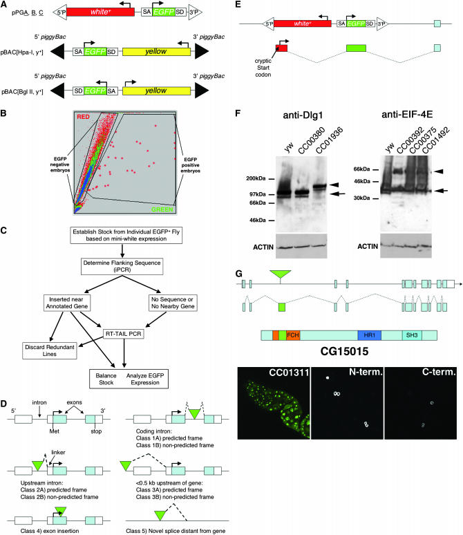
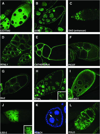
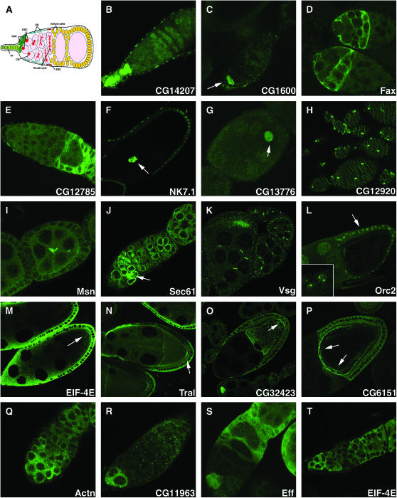
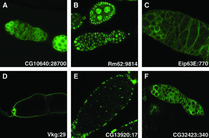
Metazoan physiology depends on intricate patterns of gene expression that remain poorly known. Using transposon mutagenesis in Drosophila, we constructed a library of 7404 protein trap and enhancer trap lines, the Carnegie collection, to facilitate gene expression mapping at single-cell resolution. By sequencing the genomic insertion sites, determining splicing patterns downstream of the enhanced green fluorescent protein (EGFP) exon, and analyzing expression patterns in the ovary and salivary gland, we found that 600-900 different genes are trapped in our collection. A core set of 244 lines trapped different identifiable protein isoforms, while insertions likely to act as GFP-enhancer traps were found in 256 additional genes. At least 8 novel genes were also identified. Our results demonstrate that the Carnegie collection will be useful as a discovery tool in diverse areas of cell and developmental biology and suggest new strategies for greatly increasing the coverage of the Drosophila proteome with protein trap insertions.


Figures

F<sc>igure</sc> 1.—
Figure 1.—
Generation and classification of protein trap vector insertions. (A) Schematic of protein trap vectors (after Morin et al. 2001). (B) Sample output from automated sorting of Drosophila embryos mobilized from site not expressing GFP. Rare GFP+ embryos (red circles) registering above a threshold value are diverted by the machine and later used to start individual cultures. (C) Scheme for characterization of putative protein trap lines (see text). (D) Classification of the general types of relationships between transposon inserts and the local genome annotation. Classes 1–4 consist of insertions in the appropriate orientation located within a codon intron (class 1), a noncoding transcribed region (class 2), an upstream genomic region (class 3), or an exon (class 4). For each class, the insert was either of the appropriate frame (subclass A) or of nonappropriate frame (subclass B) to fuse to the protein if splicing continued to the next annotated exon splice acceptor site. Class 5 consists of transposons inserted >0.5 kb from a correctly oriented annotated gene. (E) The structure of cryptic transcripts initiated within the Drosophila mini-white marker gene that contain an ATG codon and splice in frame to EGFP, thereby allowing expression independent of an endogenous transcript in some lines. (F) Western blot analysis of Dlg1 and eIF-4E protein production in control animals (y w, CC00380) and insertion lines predicted to trap Dlg1 (CC01936) or eIF-4E (CC00392, CC00375, and CC01492). (G) Abnormal nuclear accumulation of CG15015-EGFP in line CC01311 whose insertion lies within the FHC domain (left). Tissue culture cells expressing N-terminal or C-terminal fusions are found in the cytoplasm (center and right).
F<sc>igure</sc> 2.—
Figure 2.—
Protein traps for the study of protein subcellular localization. Patterns of subcellular localization of EGFP expressed from the following lines that trap the indicated genes were observed: (A) cytoplasmic, CA06607 (CG17342); (B) nuclear, BA00164 (dom); (C) enhancer trap nuclear, CB04353 (Dad) in stem cells and early cystocytes; (D) endoplasmic reticulum, CA06523 (Rtnl1); (E) extracellular, CA06735 (cathepsin K); (F) membrane, CA07474 (Picot); (G) apical, CC01941 (Baz); (H) chromatin, CA07249 (stwl); (I) nuclear membrane, CA07301 (Fs(2)Ket); (J) lipid droplets, CA07051 (Lsd-2); (K) novel structure, CA07332 (CG6854); (L) novel structure, CC01326 (polo).
F<sc>igure</sc> 3.—
Figure 3.—
Protein traps for the study of developmental regulation during oogenesis. The expression in the ovary of various protein trap lines is shown to illustrate how they can be used to associate genes with developmental processes. (A) Schematic of an ovariole tip. The terminal filament (TF), cap cells (CpC), germline stem cells (GSC), cystoblast (CB), and escort cells (ES) are illustrated. (B–H) Cell type identification. (B) Terminal filament, CB02069 (CG14207); (C) cap cells, CB03410 (CG1600); (D) escort cells, CC01359 (fax); (E) follicle cells, CC06135 (CG12785); (F) outer border cells and posterior follicle cells, CB02349 (NK7.1); (G) oocyte nucleus equals the germinal vesicle (arrow), CB04219 (CG13776); (H) novel sheath cell type, CC01646 (CG12920). (I–L) Analyzing developmental processes. (I) Novel structure in center of midstage follicle, CC00523 (Msn); (J) fusome, CC01436 (Sec61); (K) germline and somatic ring canals, CA07004 (Vsg); (L) chorion gene amplification, CB04400 (Orc2). (M–P) Localization of proteins in the oocyte. (M) Posterior pole, CC01442 (EIF-4E); (N) posterior pole, CA06517 (Tral); (O) posterior pole, CC00236 (CG32423); (P) anterior pole, CA07529 (CG6151). (Q–T) Developmental regulation of gene expression in early germ cells. (Q) Control with little change, CC01961 (Actn); (R) GSC/CB enriched, CC06238 (CG11963); (S) GSC and early cyst enriched, CC01915 (Eff); (T) GSC and forming cyst enriched, CC01442 (EIF-4E).
F<sc>igure</sc> 4.—
Figure 4.—
Studies of protein trap expression We compared the apparent intensity of GFP-protein staining in the GSCs with the RNA level of the corresponding gene as determined by Affymetrix arrays (Kai et al. 2005). (A–F) The pattern of protein trap expression of the indicated gene (see Table 4 for strain names). The expression level from Affymetrix software (mean of three measurements) is given.

Similar articles

Cited by

References

    1. Arensburger, P., Y. J. Kim, J. Orsetti, C. Aluvihare, D. A. O'Brochta et al., 2005. An active transposable element, Herves, from the African malaria mosquito Anopheles gambiae. Genetics 169: 697–708. - PMC - PubMed
    1. Aspenstrom, P., 1997. A Cdc42 target protein with homology to the non-kinase domain of FER has a potential role in regulating the actin cytoskeleton. Curr. Biol. 7: 479–487. - PubMed
    1. Bellen, H. J., R. W. Levis, G. Liao, Y. He, J. W. Carlson et al., 2004. The BDGP gene disruption project: single transposon insertions associated with 40% of Drosophila genes. Genetics 167: 761–781. - PMC - PubMed
    1. Bonin, C. P., and R. S. Mann, 2004. A piggyBac transposon gene trap for the analysis of gene expression and function in Drosophila. Genetics 167: 1801–1811. - PMC - PubMed
    1. Buszczak, M., and A. C. Spradling, 2006. The Drosophila P68 RNA helicase regulates transcriptional deactivation by promoting RNA release from chromatin. Genes Dev. 20: 977–989. - PMC - PubMed

Publication types

MeSH terms

LinkOut - more resources