Rmd9p controls the processing/stability of mitochondrial mRNAs and its overexpression compensates for a partial deficiency of oxa1p in Saccharomyces cerevisiae
- PMID: 17194787
- PMCID: PMC1840076
- DOI: 10.1534/genetics.106.063883
Rmd9p controls the processing/stability of mitochondrial mRNAs and its overexpression compensates for a partial deficiency of oxa1p in Saccharomyces cerevisiae
Abstract
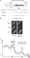
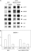
Oxa1p is a key component of the general membrane insertion machinery of eukaryotic respiratory complex subunits encoded by the mitochondrial genome. In this study, we have generated a respiratory-deficient mutant, oxa1-E65G-F229S, that contains two substitutions in the predicted intermembrane space domain of Oxa1p. The respiratory deficiency due to this mutation is compensated for by overexpressing RMD9. We show that Rmd9p is an extrinsic membrane protein facing the matrix side of the mitochondrial inner membrane. Its deletion leads to a pleiotropic effect on respiratory complex biogenesis. The steady-state level of all the mitochondrial mRNAs encoding respiratory complex subunits is strongly reduced in the Deltarmd9 mutant, and there is a slight decrease in the accumulation of two RNAs encoding components of the small subunit of the mitochondrial ribosome. Overexpressing RMD9 leads to an increase in the steady-state level of mitochondrial RNAs, and we discuss how this increase could suppress the oxa1 mutations and compensate for the membrane insertion defect of the subunits encoded by these mRNAs.
Figures


Similar articles
-
The transcriptional activator HAP4 is a high copy suppressor of an oxa1 yeast mutation.Gene. 2005 Jul 18;354:53-7. doi: 10.1016/j.gene.2005.03.016. Gene. 2005. PMID: 15908145
-
A mutational analysis reveals new functional interactions between domains of the Oxa1 protein in Saccharomyces cerevisiae.Mol Microbiol. 2010 Jan;75(2):474-88. doi: 10.1111/j.1365-2958.2009.07001.x. Epub 2009 Dec 16. Mol Microbiol. 2010. PMID: 20025673
-
Multiple defects in the respiratory chain lead to the repression of genes encoding components of the respiratory chain and TCA cycle enzymes.J Mol Biol. 2009 Apr 17;387(5):1081-91. doi: 10.1016/j.jmb.2009.02.039. Epub 2009 Feb 23. J Mol Biol. 2009. PMID: 19245817
-
The enigmatic role of Mim1 in mitochondrial biogenesis.Eur J Cell Biol. 2010 Feb-Mar;89(2-3):212-5. doi: 10.1016/j.ejcb.2009.11.002. Epub 2009 Nov 26. Eur J Cell Biol. 2010. PMID: 19944477 Review.
-
Protein insertion into the inner membrane of mitochondria.IUBMB Life. 2003 Apr-May;55(4-5):219-25. doi: 10.1080/1521654031000123349. IUBMB Life. 2003. PMID: 12880202 Review.
Cited by
-
Genomewide Elucidation of Drug Resistance Mechanisms for Systemically Used Antifungal Drugs Amphotericin B, Caspofungin, and Voriconazole in the Budding Yeast.Antimicrob Agents Chemother. 2019 Aug 23;63(9):e02268-18. doi: 10.1128/AAC.02268-18. Print 2019 Sep. Antimicrob Agents Chemother. 2019. PMID: 31209012 Free PMC article.
-
Mitochondrial protein synthesis, import, and assembly.Genetics. 2012 Dec;192(4):1203-34. doi: 10.1534/genetics.112.141267. Genetics. 2012. PMID: 23212899 Free PMC article.
-
The ribosome receptors Mrx15 and Mba1 jointly organize cotranslational insertion and protein biogenesis in mitochondria.Mol Biol Cell. 2018 Oct 1;29(20):2386-2396. doi: 10.1091/mbc.E18-04-0227. Epub 2018 Aug 9. Mol Biol Cell. 2018. PMID: 30091672 Free PMC article.
-
Translation initiation in Saccharomyces cerevisiae mitochondria: functional interactions among mitochondrial ribosomal protein Rsm28p, initiation factor 2, methionyl-tRNA-formyltransferase and novel protein Rmd9p.Genetics. 2007 Mar;175(3):1117-26. doi: 10.1534/genetics.106.064576. Epub 2006 Dec 28. Genetics. 2007. PMID: 17194786 Free PMC article.
-
Cbp3-Cbp6 interacts with the yeast mitochondrial ribosomal tunnel exit and promotes cytochrome b synthesis and assembly.J Cell Biol. 2011 Jun 13;193(6):1101-14. doi: 10.1083/jcb.201103132. J Cell Biol. 2011. PMID: 21670217 Free PMC article.
References
-
- Altamura, N., N. Capitanio, N. Bonnefoy, S. Papa and G. Dujardin, 1996. The Saccharomyces cerevisiae OXA1 gene is required for the correct assembly of cytochrome c oxidase and oligomycin-sensitive ATP synthase. FEBS Lett. 382: 111–115. - PubMed
-
- Amiott, E., and J. Jaehning, 2006. Mitochondrial transcription is regulated via an ATP sensing mechanism that couples RNA abundance to respiration. Mol. Cell 22: 329–338. - PubMed
-
- Barrientos, A., M. H. Barros, I. Valnot, A. Rotig, P. Rustin et al., 2002. Cytochrome oxidase in health and disease. Gene 286: 53–63. - PubMed
-
- Bauer, M., M. Behrens, K. Esser, G. Michaelis and E. Pratje, 1994. PET1402, a nuclear gene required for proteolytic processing of cytochrome oxidase subunit 2 in yeast. Mol. Gen. Genet. 245: 272–278. - PubMed
Publication types
MeSH terms
Substances
LinkOut - more resources
Full Text Sources
Molecular Biology Databases

