. 2007 Mar;189(6):2443-59.
doi: 10.1128/JB.01688-06. Epub 2006 Dec 28.

Diversity of the abundant pKLC102/PAGI-2 family of genomic islands in Pseudomonas aeruginosa

Affiliations

Diversity of the abundant pKLC102/PAGI-2 family of genomic islands in Pseudomonas aeruginosa

Jens Klockgether et al. J Bacteriol. 2007 Mar.

Abstract

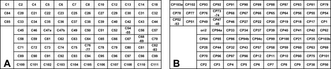
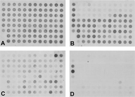
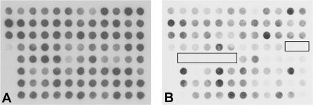
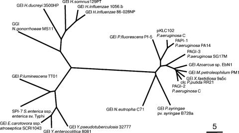
The known genomic islands of Pseudomonas aeruginosa clone C strains are integrated into tRNA(Lys) (pKLC102) or tRNA(Gly) (PAGI-2 and PAGI-3) genes and differ from their core genomes by distinctive tetranucleotide usage patterns. pKLC102 and the related island PAPI-1 from P. aeruginosa PA14 were spontaneously mobilized from their host chromosomes at frequencies of 10% and 0.3%, making pKLC102 the most mobile genomic island known with a copy number of 30 episomal circular pKLC102 molecules per cell. The incidence of islands of the pKLC102/PAGI-2 type was investigated in 71 unrelated P. aeruginosa strains from diverse habitats and geographic origins. pKLC102- and PAGI-2-like islands were identified in 50 and 31 strains, respectively, and 15 and 10 subtypes were differentiated by hybridization on pKLC102 and PAGI-2 macroarrays. The diversity of PAGI-2-type islands was mainly caused by one large block of strain-specific genes, whereas the diversity of pKLC102-type islands was primarily generated by subtype-specific combination of gene cassettes. Chromosomal loss of PAGI-2 could be documented in sequential P. aeruginosa isolates from individuals with cystic fibrosis. PAGI-2 was present in most tested Cupriavidus metallidurans and Cupriavidus campinensis isolates from polluted environments, demonstrating the spread of PAGI-2 across habitats and species barriers. The pKLC102/PAGI-2 family is prevalent in numerous beta- and gammaproteobacteria and is characterized by high asymmetry of the cDNA strands. This evolutionarily ancient family of genomic islands retained its oligonucleotide signature during horizontal spread within and among taxa.


Figures

FIG. 1.
FIG. 1.
Schematic diagram of the positions of ORF-derived PCR products on the PAGI-2 (A) and pKLC102 (B) macroarrays. (A) PAGI-2. ORF C47 is represented twice (C47a and C47b) by different PCR products. (B) pKLC102. ORF CP103 is represented twice (CP103a and CP103b) and CP94 three times (CP94a, CP94b, and CP94c) by different PCR products. Five (A) or 10 (B) positive or negative control dots were spotted in the lower left corner.
FIG. 2.
FIG. 2.
Tetranucleotide usage of the four P. aeruginosa genomic islands pKLC102, PAPI-1, PAGI-2, and PAGI-3. Local OU patterns were analyzed in 5-kb sliding windows with steps of 0.5 kb. Curves of the distance D:n0_4mer, pattern skew PS:n0_4mer, and oligonucleotide variance OUV:n1_4mer are specified by color code: blue for D, green for PS and brown for OUV. Protein-coding genes are shown by red bars. The abscissa separates genes by their direction of transcription. The tetranucleotide usage of the genomic islands was significantly different from that of the whole chromosome. The median (inner quartile) values of local tetranucleotide patterns in the whole P. aeruginosa PAO1 chromosome were 13.9 (12.3 to 16.0) for D:n0_4mer, 21.4 (17.9 to 25.6) for PS:n0_4mer, and 0.37 (0.32 to 0.43) for OUV:n1_4mer.
FIG. 3.
FIG. 3.
Combinatorial PCR analysis of integrated and episomal versions of genomic islands PAPI-1 in strain P. aeruginosa PA14 and pKLC102 in P. aeruginosa SG17M. An aliquot from an exponentially growing culture was inoculated into 100 ml fresh medium adjusted to an optical density at 578 nm (OD578) of 0.2. Samples were then taken from the growing culture (from left to right) at OD578s of 0.9, 1.3, 2.0, 2.9, and 4.0 and after 24 h (left) or at OD578s of 0.9, 1.3, 2.0, and 4.0 and after 24 h (right). Bacteria were growing aerobically in 250-ml flasks in liquid LB medium at 37°C at a mixing frequency of 250 rpm. Chromosome-integrated islands were detected by PCR products spanning the 5′ tRNA (il) or the 3′ tRNA (ir) integration sites by utilizing PA14- and PAPI-1- or SG17M- and pKLC102-derived primer sequences. Circularized episomal forms (ce) were identified by PCR products spanning the breakpoints in PAPI-1 or pKLC102. PA14 or SG17M chromosomes (fa) devoid of PAPI-1 or pKLC102 were detected by PCR products spanning the tRNALys gene adjacent to the PAO1 homolog PA4541. PCR kinetics were performed with 50 ng P. aeruginosa DNA in a 50-μl reaction mixture. Aliquots of 5 μl were withdrawn at the indicated cycles, separated by electrophoresis, and stained with ethidium bromide.
FIG. 4.
FIG. 4.
Examples of PAGI-2 (upper two rows) and pKLC102 (lower two rows) subtype macroarray hybridization patterns. The PAGI-2 macroarrays show (A) strain PAO (DSM1707) (negative control), (B) strain C (positive control), (C) strain 7 (subtype G1b), (D) strain 3 (subtype G2a), (E) strain 54 (subtype G2c), and (F) strain 63 (subtype G4). The pKLC102 macroarrays show (G) strain PAO (DSM1707) (negative control), (H) strain SG17M (positive control), (I) strain 6 (subtype K1c), (J) strain 10 (subtype K3c), (K) strain 36 (subtype K3d), and (L) strain 53 (subtype K4).
FIG. 5.
FIG. 5.
Summary of macroarray hybridization data for 31 PAGI-2-type-positive (A) and 50 pKLC102-type-positive (B) P. aeruginosa strains. The shading indicates the percentages of island-positive strains with a hybridization signal for the respective ORF. Black, ≥96% of strains positive; dark gray, 90 to 95% positive; light gray, 50 to 89% positive; white, <50% positive.
FIG. 6.
FIG. 6.
Relatedness of macroarray hybridization patterns of 55 PAGI-2- and/or pKLC102-positive P. aeruginosa strains. The unrooted tree is based on the parsimony analysis (“PHYLIP 3.66”) of the hybridization data.
FIG. 7.
FIG. 7.
Loss of PAGI-2-type islands in sequential P. aeruginosa airway isolates from patients with cystic fibrosis. (Upper row) PAGI-2 macroarray hybridization patterns of clone C strains SG1 (A) and SG3 (B), indicating the loss of PAGI-2 in the later isolate SG3 while another PAGI-2 subtype was retained. SG1 (strain C) was isolated from the patient's first P. aeruginosa-positive sputum specimen; SG3 is the sixth isolate, collected 2 years later. (Lower row) PAGI-2 macroarray hybridization patterns of clone C strains NN18 (C) and NN86 (D), indicating the loss of a PAGI-2-type island(s) in strain NN86, which was isolated from the patient′s last clone C-positive culture 17 years after the acquisition of clone C.
FIG. 8.
FIG. 8.
PAGI-2 macroarray hybridization patterns of Cupriavidus strains C. campinensis AE2701 (A) and C. metallidurans CH79 (B). The boxes highlight absent hybridization signals.
FIG. 9.
FIG. 9.
Similarity of pKLC102-type genomic islands in proteobacteria based on the distance of oligonucleotide usage. The distance D:n0_4mer of tetranucleotides was calculated for each genomic island. The matrix of D values obtained was sorted for the degree of evolutionary relationship between the genomic islands by the Fitch-Margoliash criterion, assuming a constant molecular clock, and by the least-squares methods using the KITSCH program of the PHYLIP library (11). The bar indicates branch length 5. Branch lengths are not drawn exactly to scale. Short branches are exaggerated in length so that they are more visible.
FIG. 10.
FIG. 10.
Pattern skew of pKLC102-type genomic islands (squares) and their corresponding chromosomes (triangles). Pattern skew values (n0_4mer PS) are plotted against the logarithmic scale of sequence lengths. The gray-shaded area depicts the 95% confidence intervals of variation of n0_4mer PS values in 155 completely sequenced bacterial chromosomes and 316 plasmids (37). The PS values of n0_4mer patterns of bacterial chromosomes are typically in the range of 1 to 8%. Outliers in the investigated panel are Haemophilus ducreyi 35000HP, with a PS value of about 9%, and Xylella fastidiosa 9a5c, with an extreme value of 24.3%. The n0_4mer PS values of pKLC102-type genomic islands exceed the 95% confidence interval. The genomic islands, identified by their host strains, with the name of the island given in brackets if available, were as follows: 1, Azoarcus sp. strain EbN1; 2, Erwinia carotovora subsp. atroseptica SCRI1043; 3, Haemophilus ducreyi 3500HP; 4, Haemophilus influenzae 86-028NP (ICEHin-like); 5, Haemophilus somnus 129PT; 6, Methylobium petroleophilum PM1; 7, Photorhabdus luminescens TT01; 8, P. aeruginosa C (pKLC102); 9, P. aeruginosa C (PAGI-2); 10, P. aeruginosa PA14 (PAPI-1); 11, P. aeruginosa SG17M (PAGI-3); 12, Pseudomonas fluorescens Pf-5; 13, Pseudomonas syringae pv. syringae B728a; 14, Salmonella enterica subsp. enterica serovar Typhi CT18 (SPI-7); 15, Xylella fastidiosa 9a5c; 16, Yersinia enterocolitica 8081; 17, H. influenzae 1056.b (ICEHin 1056); 18, Neisseria gonorrhoeae MS11 (GGI); 19, Nitrosomonas eutropha C71; 20, Pseudomonas putida RR21 (clc-transposon); 21, P. syringae pv. phaseolicola 1302A (PPHG-1); 22, Yersinia pseudotuberculosis 32777 (YAPI).

Similar articles

Cited by

References

    1. Almagor, H. 1983. A Markov analysis of DNA sequences. J. Theor. Biol. 104:633-645. - PubMed
    1. Ausubel, F. M., R. Brent, R. E. Kingston, D. D. Moore, J. G. Seidmann, J. A. Smith, and K. Struhl, ed. 1994. Current protocols in molecular biology. Wiley, New York, NY.
    1. Baisnee, P. F., S. Hampson, and P. Baldi. 2002. Why are complementary DNA strands symmetric? Bioinformatics 18:1021-1033. - PubMed
    1. Baldi, P., and P. F. Baisnee. 2000. Sequence analysis by additive scales: DNA structure for sequences and repeats of all lengths. Bioinformatics 16:865-889. - PubMed
    1. Bragonzi, A., L. Wiehlmann, J. Klockgether, N. Cramer, D. Worlitzsch, G. Döring, and B. Tümmler. 2006. Sequence diversity of the mucABD locus in Pseudomonas aeruginosa isolates from patients with cystic fibrosis. Microbiology 152:3261-3269. - PubMed

Publication types

MeSH terms

LinkOut - more resources