

"Candidatus Helicobacter heilmannii" from a cynomolgus monkey induces gastric mucosa-associated lymphoid tissue lymphomas in C57BL/6 mice
- PMID: 17194807
- PMCID: PMC1828597
- DOI: 10.1128/IAI.01459-06
"Candidatus Helicobacter heilmannii" from a cynomolgus monkey induces gastric mucosa-associated lymphoid tissue lymphomas in C57BL/6 mice
Abstract
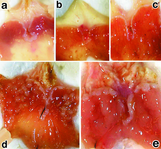
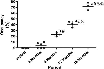
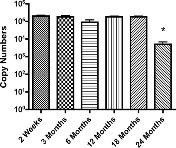
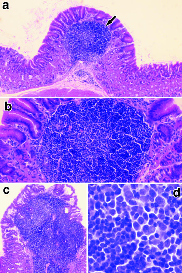
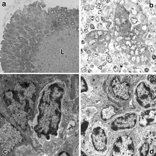
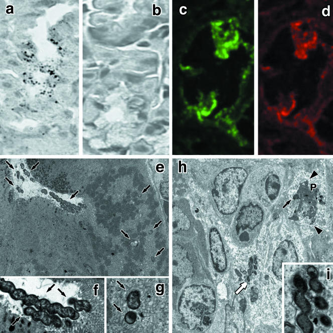
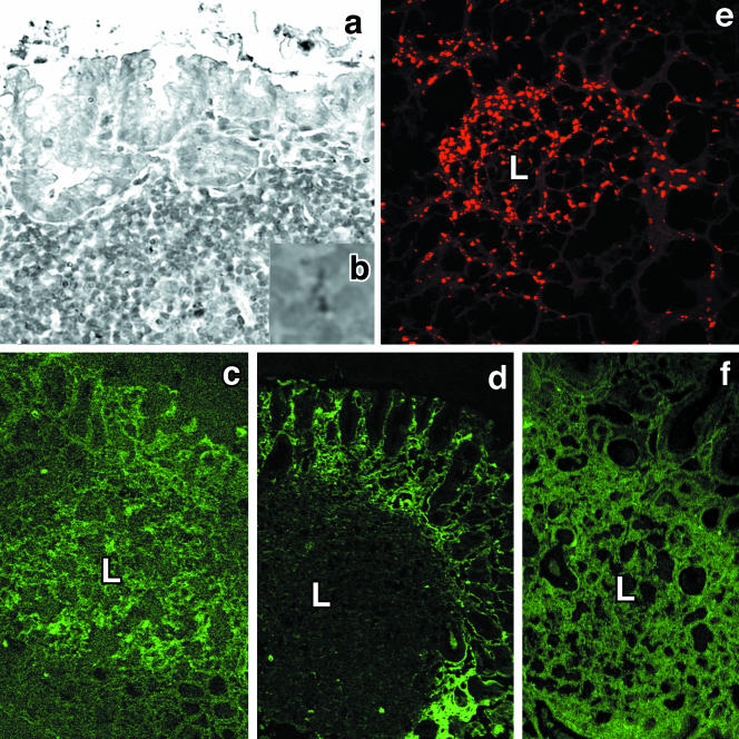
Both Helicobacter pylori and "Candidatus Helicobacter heilmannii" infections are associated with peptic ulcers, gastric adenocarcinoma, and gastric mucosa-associated lymphoid tissue (MALT) lymphomas. However, good animal models of H. pylori clinical diseases are rare. In this study, we aimed to establish an animal model of "Candidatus Helicobacter heilmannii" gastric MALT lymphoma. We used a urease-positive gastric mucosal and mucus homogenate from a cynomolgus monkey maintained in C57BL/6 mouse stomachs. The bacterium in the homogenate was identified as "Candidatus Helicobacter heilmannii" based on a DNA sequence analysis of the 16S rRNA and urease genes. Mucosal and mucus homogenates were used to inoculate C57BL/6 mice, which were then examined for 24 months. We observed a gradual increase in the surface area of protrusive lesions in almost all infected C57BL/6 mouse fundic stomachs 6 months after infection. Light microscopic observations revealed an accumulation of B lymphocytes along with destruction of glandular elements and the presence of lymphoepithelial lesions consistent with low-grade MALT lymphomas. Electron microscopic observation revealed numerous "Candidatus Helicobacter heilmannii" bacilli in the fundic glandular lumen, the intracellular canaliculi, and the cytoplasm of intact cells, as well as damaged parietal cells. In conclusion, "Candidatus Helicobacter heilmannii" induced gastric MALT lymphomas in almost 100% of infected C57BL/6 mice after a 6-month period associated with the destruction of parietal cells.
Figures
Similar articles
-
Increased apoptosis and angiogenesis in gastric low-grade mucosa-associated lymphoid tissue-type lymphoma by Helicobacter heilmannii infection in C57/BL6 mice.FEMS Immunol Med Microbiol. 2007 Jul;50(2):268-72. doi: 10.1111/j.1574-695X.2007.00252.x. Epub 2007 May 4. FEMS Immunol Med Microbiol. 2007. PMID: 17488330
-
Gastric B-cell mucosa-associated lymphoid tissue (MALT) lymphoma in an animal model of 'Helicobacter heilmannii' infection.J Pathol. 2004 Aug;203(4):896-903. doi: 10.1002/path.1593. J Pathol. 2004. PMID: 15258991
-
Microcirculatory alteration in low-grade gastric mucosa-associated lymphoma by Helicobacter heilmannii infection: its relation to vascular endothelial growth factor and cyclooxygenase-2.J Gastroenterol Hepatol. 2008 Dec;23 Suppl 2:S157-60. doi: 10.1111/j.1440-1746.2008.05554.x. J Gastroenterol Hepatol. 2008. PMID: 19120890
-
[Relation of Helicobacter heilmannii to gastric mucosal damage as a model of zoonosis between men and pets].Nihon Rinsho. 2005 Nov;63 Suppl 11:605-8. Nihon Rinsho. 2005. PMID: 16363610 Review. Japanese. No abstract available.
-
Gastric mucosa-associated lymphoid tissue lymphoma: implications of animal models on pathogenic and therapeutic considerations--mouse models of gastric lymphoma.Recent Results Cancer Res. 2000;156:42-51. doi: 10.1007/978-3-642-57054-4_6. Recent Results Cancer Res. 2000. PMID: 10802862 Review.
Cited by
-
Helicobacter suis causes severe gastric pathology in mouse and mongolian gerbil models of human gastric disease.PLoS One. 2010 Nov 22;5(11):e14083. doi: 10.1371/journal.pone.0014083. PLoS One. 2010. PMID: 21124878 Free PMC article.
-
Involvement of non-Helicobacter pylori helicobacter infections in Helicobacter pylori-negative gastric MALT lymphoma pathogenesis and efficacy of eradication therapy.Gastric Cancer. 2021 Jul;24(4):937-945. doi: 10.1007/s10120-021-01172-x. Epub 2021 Feb 27. Gastric Cancer. 2021. PMID: 33638751
-
Overexpression of miR-142-5p and miR-155 in gastric mucosa-associated lymphoid tissue (MALT) lymphoma resistant to Helicobacter pylori eradication.PLoS One. 2012;7(11):e47396. doi: 10.1371/journal.pone.0047396. Epub 2012 Nov 28. PLoS One. 2012. PMID: 23209550 Free PMC article.
-
A case of Helicobacter heilmannii-associated primary gastric mucosa-associated lymphoid tissue lymphoma achieving complete remission after eradication.Clin J Gastroenterol. 2013 Feb;6(1):38-45. doi: 10.1007/s12328-012-0355-9. Epub 2013 Jan 9. Clin J Gastroenterol. 2013. PMID: 26181403
-
Chronic inflammatory disease, lymphoid tissue neogenesis and extranodal marginal zone B-cell lymphomas.Haematologica. 2009 Aug;94(8):1109-23. doi: 10.3324/haematol.2009.005983. Epub 2009 Jul 16. Haematologica. 2009. PMID: 19608670 Free PMC article. Review.
References
-
- Andersen, L. P., A. Norgaard, S. Holck, J. Blom, and T. L. Elsborg. 1996. Isolation of a “Helicobacter heilmanii”-like organism from the human stomach. Eur. J. Clin. Microbiol. Infect. Dis. 15:95-96. - PubMed
-
- Chisholm, S. A., and R. J. Owen. 2003. Development and application of a novel screening PCR assay for direct detection of ‘Helicobacter heilmannii’-like organisms in human gastric biopsies in southeast England. Diagn. Microbiol. Infect. Dis. 46:1-7. - PubMed
-
- Dubois, A., L. M. Heman-Ackah, E. S. Drazek, A. Tarnawski, W. N. Fishbein, G. I. Perez-Perez, and M. J. Blazer. 1994. Natural gastric infection with Helicobacter pylori in monkeys: a model for spiral bacterial infection in humans. Gastroenterology 160:1405-1417. - PubMed
Publication types
MeSH terms
LinkOut - more resources
Full Text Sources
Other Literature Sources
Medical
Molecular Biology Databases
