

Binding of antiphospholipid antibodies to discontinuous epitopes on domain I of human beta(2)-glycoprotein I: mutation studies including residues R39 to R43
- PMID: 17195232
- PMCID: PMC2117024
- DOI: 10.1002/art.22306
Binding of antiphospholipid antibodies to discontinuous epitopes on domain I of human beta(2)-glycoprotein I: mutation studies including residues R39 to R43
Abstract
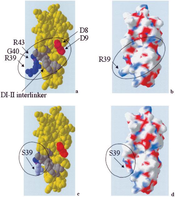
Objective: Pathogenic antiphospholipid antibodies (aPL) bind the self antigen N-terminal domain (domain I) of beta(2)-glycoprotein I (beta(2)GPI), with residues G40-R43 being important. However, peptides homologous to other regions of domain I have also been shown to bind aPL. Furthermore, there are no published reports of the effects of altering R39, which has greater surface exposure than the G40-R43 residues.
Methods: We used a novel, efficient method of production and purification of human domain I by Escherichia coli to create multiple mutants of domain I. These domain I mutants were then screened for binding to a range of polyclonal IgG purified from patients with antiphospholipid syndrome, using both solid-phase and fluid-phase assays.
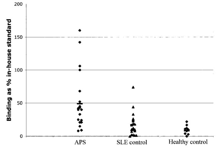
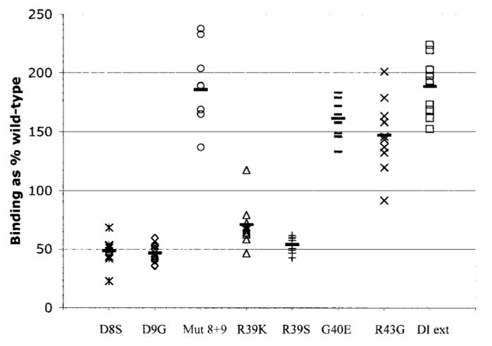
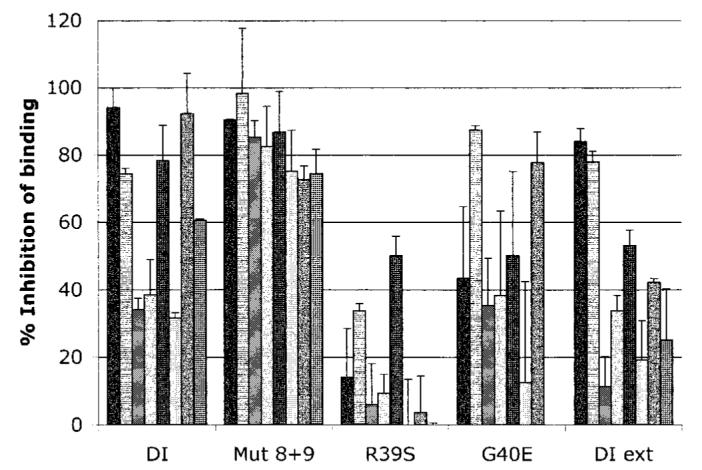
Results: E coli-expressed purified domain I selectively bound IgG derived from patients with antiphospholipid syndrome. In region R39-R43, the R39S mutation had the greatest effect in terms of reducing binding to a panel of aPL in the fluid phase (mean +/- SD inhibition 14 +/- 18.5% versus 44.1 +/- 31.7% for G40E and 62.9 +/- 25.7% for wild-type domain I). Conversely, altering both D8 and D9 to S8 and G9, respectively, had the effect of enhancing binding to aPL in the fluid phase. Adding the remainder of the domain I-II interlinker resulted in enhanced binding over wild-type in the solid phase but not the fluid phase.
Conclusion: The binding of aPL to beta(2)GPI domain I is complex and likely to involve discontinuous epitopes that include R39 in addition to G40-R43, the domain I-II interlinker, and possibly D8 and D9. Domain I variants with enhanced binding to aPL compared with wild-type domain I may aid in the development of novel therapies.
Figures
References
-
- Miyakis S, Lockshin MD, Atsumi T, Branch DW, Brey RL, Cervera R, et al. International consensus statement on an update of the classification criteria for definite antiphospholipid syndrome. J Thromb Haemost. 2006;4:295–306. - PubMed
-
- Pierangeli SS, Harris EN. In vivo models of thrombosis for the antiphospholipid syndrome. Lupus. 1996;5:451–5. - PubMed
-
- Pierangeli SS, Liu X, Espinola R, Olee T, Zhu M, Harris NE, et al. Functional analyses of patient-derived IgG monoclonal anticardiolipin antibodies using in vivo thrombosis and in vivo microcirculation models. Thromb Haemost. 2000;84:388–95. - PubMed
-
- Raschi E, Testoni C, Bosisio D, Borghi MO, Koike T, Mantovan A, et al. Role of the MyD88 transduction signaling pathway in endothelial activation by antiphospholipid antibodies. Blood. 2003;101:3495–500. - PubMed
-
- Pierangeli SS, Colden-Stanfield M, Liu X, Barker JH, Anderson GL, Harris EN. Antiphospholipid antibodies from antiphospholipid syndrome patients activate endothelial cells in vitro and in vivo. Circulation. 1999;99:1997–2002. - PubMed
Publication types
MeSH terms
Substances
Grants and funding
LinkOut - more resources
Full Text Sources
Other Literature Sources
Molecular Biology Databases
