Case Reports
. 2007 Mar;46(3):492-8.
doi: 10.1016/j.jhep.2006.09.022. Epub 2006 Nov 9.

Glycogen storage disease type III-hepatocellular carcinoma a long-term complication?

Affiliations
Case Reports

Glycogen storage disease type III-hepatocellular carcinoma a long-term complication?

Erin Demo et al. J Hepatol. 2007 Mar.

Abstract

Background/aims: Glycogen storage disease III (GSD III) is caused by a deficiency of glycogen-debranching enzyme which causes an incomplete glycogenolysis resulting in glycogen accumulation with abnormal structure (short outer chains resembling limit dextrin) in liver and muscle. Hepatic involvement is considered mild, self-limiting and improves with age. With increased survival, a few cases of liver cirrhosis and hepatocellular carcinoma (HCC) have been reported.

Methods: A systematic review of 45 cases of GSD III at our center (20 months to 67 years of age) was reviewed for HCC, 2 patients were identified. A literature review of HCC in GSD III was performed and findings compared to our patients.

Conclusions: GSD III patients are at risk for developing HCC. Cirrhosis was present in all cases and appears to be responsible for HCC transformation There are no reliable biomarkers to monitor for HCC in GSD III. Systematic evaluation of liver disease needs be continued in all patients, despite lack of symptoms. Development of guidelines to allow for systematic review and microarray studies are needed to better delineate the etiology of the hepatocellular carcinoma in patients with GSD III.


Figures

Fig. 1
Fig. 1
Patient 1. (A) A 54-year-old male with GSD type IIIa and hepatocellular carcinoma. (A) IV contrast enhanced CT examination of the upper liver shows heterogeneous parenchyma with multiple low attenuation regions. The liver was not enlarged. Ascites (*) surrounds the liver. (B) Image from a IV contrast enhanced CT examination at the region of the upper porta hepatis again shows scattered low-attenuation lesions with a more focally defined low-attenuation region in the area of the left portal vein (arrows). Ascites is again evident around the liver.
Fig. 2
Fig. 2
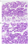
Patient 1. A fine needle aspiration cytology of one of the liver lesions (A) reveals a cellular population of tumor cells, some organized in pseudoacinar rosettes and some in trabeculae with poor cohesion and no intervening desmoplastic stroma. This architecture is characteristic of hepatocellular carcinoma in biopsy specimens. A higher power view (B) demonstrates rudimentary hepatocellular features including polygonal cells with granular eosinophilic cytoplasm and central nuclei, but with the high nuclear cytoplasmic ratio and mild nuclear irregularity of moderately well-differentiated hepatocellular carcinoma. (Fig. 1A 20× and B 40×). [This figure appears in colour on the web.]
Fig. 3
Fig. 3
Sixty-two year-old male (A) axial image of the upper abdomen from a non-contrast enhanced CT examination shows a low-attenuation lesion in the right lobe of the liver (the second lesion is not shown) (arrow). (B) IV contrast-enhanced axial CT image at the level of the portal vein performed at age 67 years shows a large, poorly defined heterogeneous mass in the same region (large arrows). A smaller area seen adjacent to the caudate lobe (small arrow) represents metastatic adenopathy. (C) Axial image from a non-contrast enhanced CT examination obtained in the left lateral decubital position at a level just inferior to that in (B) shows the low attenuation lesion in the right lobe of the liver (arrows). Fine needle aspiration of the liver was not diagnostic for malignancy. Biopsy of the adrenal lesion (large arrow) was positive for hepatocellular carcinoma.
Fig. 4
Fig. 4
Patient 2. A CT-guided biopsy of the liver reveals cirrhosis (A). Trichrome stain reveals fibrous septa surrounding regenerative nodules of hepatocytes. Hepatocytes are expanded by glycogen in the H&E stain (B) (Fig. 2A 2× and B 20×). [This figure appears in colour on the web.]
Fig. 5
Fig. 5
Patient 2. CT-guided biopsy of the adrenal mass confirms the diagnosis of metastatic well-differentiated hepatocellular carcinoma involving the adrenal gland (A) single line, adrenal and fat; double line, hepatocellular carcinoma. Similar to the carcinoma in patient 1, poorly cohesive trabeculae and pseudoacini of tumor cells are seen with granular cytoplasm, high nuclear-to-cytoplasmic ratio and mild nuclear irregularity (B) (Fig. 3A 4× and B 40×). [This figure appears in colour on the web.]

Comment in

References

    1. Chen YT, Burhchell A. Glycogen storage diseases. In: Scriver CR, Beaudet AL, Sly WS, Valle D, editors. The metabolic and molecular bases of inherited disease. New York: McGraw-Hill, Inc., Health Professions Division; pp. 935–966.
    1. Talente GM, Coleman RA, Alter C, Baker L, Brown BI, Cannon RA, et al. Glycogen storage disease in adults. Ann Intern Med. 1994;120:218–226. - PubMed
    1. Coleman RA, Winter HS, Wolf B, Chen YT. Glycogen debranching enzyme deficiency: long-term study of serum enzyme activities and clinical features. J Inherit Metab Dis. 1992;15:869–881. - PubMed
    1. Labrune P, Trioche P, Duvaltier I, Chevalier P, Odievre M. Hepatocellular adenomas in glycogen storage disease type I and III: a series of 43 patients and review of the literature. J Pediatr Gastroenterol Nutr. 1997;24:276–279. - PubMed
    1. Matern D, Starzl TE, Arnaout W, Barnard J, Bynon JS, Dhawan A, et al. Liver transplantation for glycogen storage disease types I, III, and IV. Eur J Pediatr. 1999;158 Suppl. 2:S43–S48. - PMC - PubMed

Publication types

MeSH terms