. 2007 Jan;14(1):97-105.
doi: 10.1016/j.chembiol.2006.12.005. Epub 2006 Dec 28.

Structural investigation of the GlmS ribozyme bound to Its catalytic cofactor

Affiliations

Structural investigation of the GlmS ribozyme bound to Its catalytic cofactor

Jesse C Cochrane et al. Chem Biol. 2007 Jan.

Abstract

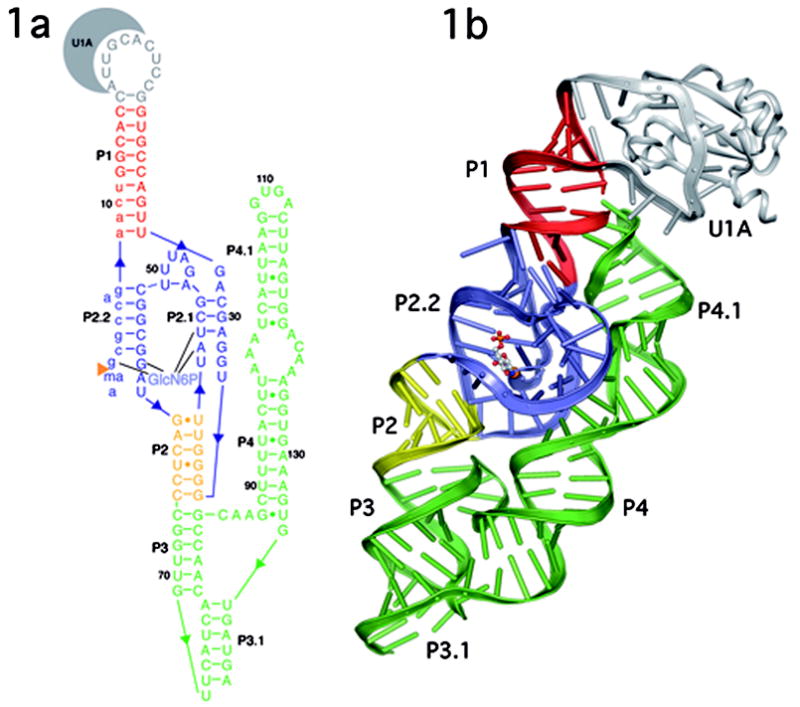
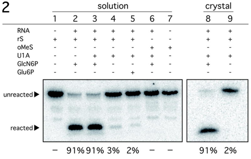
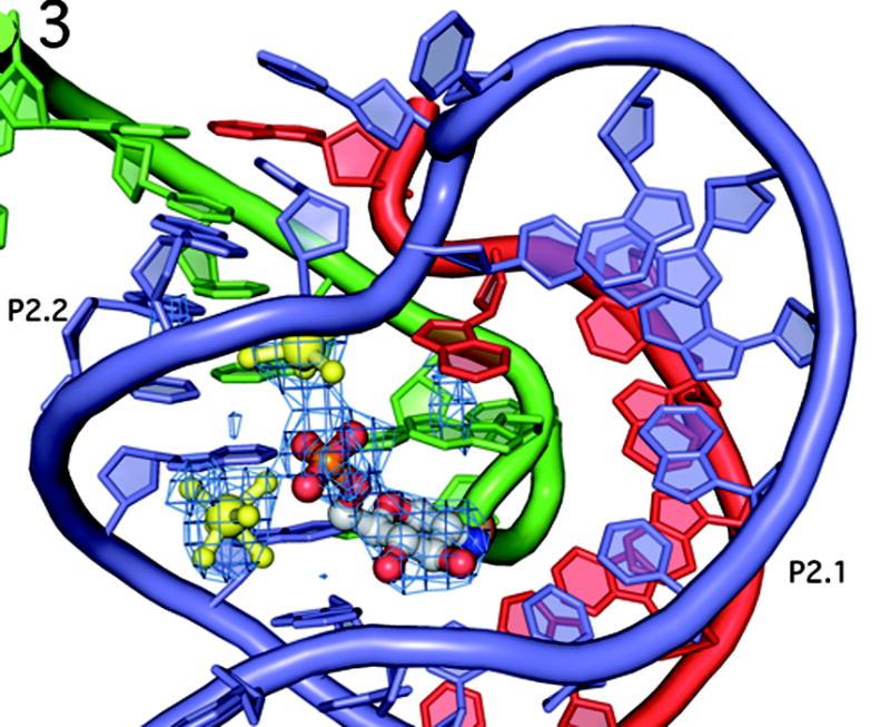
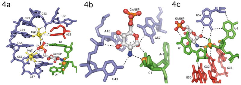
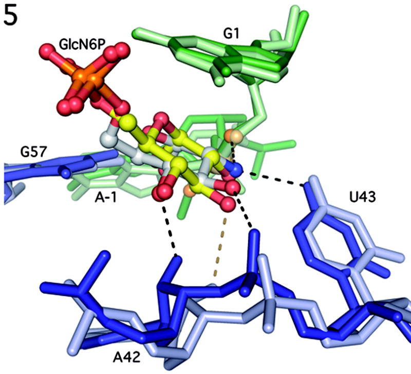
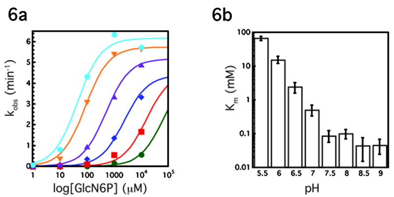
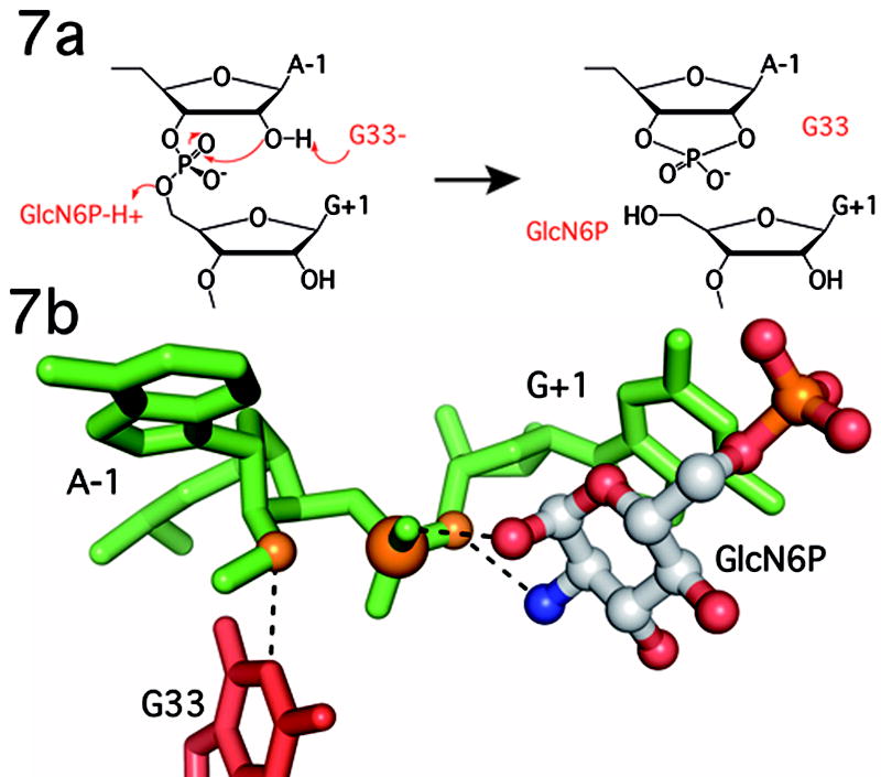
The GlmS riboswitch is located in the 5'-untranslated region of the gene encoding glucosamine-6-phosphate (GlcN6P) synthetase. The GlmS riboswitch is a ribozyme with activity triggered by binding of the metabolite GlcN6P. Presented here is the structure of the GlmS ribozyme (2.5 A resolution) with GlcN6P bound in the active site. The GlmS ribozyme adopts a compact double pseudoknot tertiary structure, with two closely packed helical stacks. Recognition of GlcN6P is achieved through coordination of the phosphate moiety by two hydrated magnesium ions as well as specific nucleobase contacts to the GlcN6P sugar ring. Comparison of this activator bound and the previously published apoenzyme complex supports a model in which GlcN6P does not induce a conformational change in the RNA, as is typical of other riboswitches, but instead functions as a catalytic cofactor for the reaction. This demonstrates that RNA, like protein enzymes, can employ the chemical diversity of small molecules to promote catalytic activity.


Figures

Figure 1
Figure 1
A. Secondary and B. Tertiary structure of the B. anthracis GlmS ribozyme depicted in ribbons format. The P1 helix is shown in red, P2 in yellow, and P3–P4 in green. The double pseudoknot, consisting of helices P2.1, P2.2, P2a and related joiner regions is in blue. GlcN6P is bound in the active site and is shown in ball and stick representation primarily in gray. U1A and the U1A binding loop are in gray.
Figure 2
Figure 2
Reactivity of the B. anthracis GlmS ribozyme construct under crystallization conditions. Activity assays conditions were similar to those used for the refolding of the GlmS RNA with a radiolabeled synthetic oligonucleotide substrate. The substrate rS has a ribose at position A-1; oMeS has a 2′-O-methyl at A-1. Reactions were performed either in solution or in crystals as indicated. The reagents included in each lane are as indicated above in lane. The mobility of the substrate and product are marked with arrows. The percent cleavage for each reaction after a 2 hour incubation is listed below the autoradiogram.
Figure 3
Figure 3
The double pseudoknotted active site of the GlmS ribozyme in the same orientation as in Fig. 1b. The color scheme has been altered to emphasize the contribution of each strand in the active site. The substrate strand (nucleotides −2 to 9) are shown in green, with the scissile phosphate depicted as an orange ball. Nucleotides 27–34 are in red, and nucleotides 41–59, which comprise the P2 loop, are in blue. This color scheme is also used in Figures 4, 5 and 7. The P2.1 pseudoknot is formed from the red and blue strands; the P2.2 pseudoknot is formed from the green and blue strands. GlcN6P is shown primarily in gray and the two hydrated magnesium ions are in yellow. Fo − Fc density is shown in blue mesh, contoured at 2.5σ, calculated before addition of GlcN6P, metal ions or water molecules to the model.
Figure 4
Figure 4
GlcN6P binding by the GlmS ribozyome. A. Phosphate coordination by two magnesium ions in the GlmS ribozyme. Nucleotides in P2.1 (blue), A28 (red) and G1 (green) use water mediated contacts to organize two hydrated magnesium ions (yellow) and orient the GlcN6P (primarily in gray) phosphate oxygens in the active site. B. Recognition of the GlcN6P sugar ring by nucleobase functional groups. The sugar contacts nucleotides A42, U43 and G57 (blue), and the sugar and 3′-phosphate of G1 (green). The guanine base at G1, which stacks on top of GlcN6P (see Fig. 4a), is not shown to allow all hydrogen bonding contacts to be visualized. The scissile phosphate, 5′-O leaving group and 2′-O nucleophile are shown as orange spheres. C. Active site interactions expected to stabilize this conformation are depicted as thin dashed lines. The catalytically critical interactions between the ethanolamine moiety of GlcN6P and the reactive phosphate are shown as thicker dashed lines. The coloring of individual nucleotides follows the convention in Fig. 3. The scissile phosphate, the 5′-O leaving group and the 2′-O nucleophile (which has been methylated) are shown as orange spheres.
Figure 5
Figure 5
Comparison of the active sites of GlcN6P bound B. anthracis GlmS ribozyme and Glu6P bound T. tengcongenis GlmS ribozyme. B. anthracis nucleotides are in darker shades of blue and green and T. tengcongenis nucleotides are in lighter shades. GlcN6P is shown primarily in gray and Glu6P is shown primarily in yellow. Hydrogen bonds to GlcN6P are show in black, hydrogen bonds to Glu6P are shown in dark yellow.
Figure 6
Figure 6
pH dependence of the Glms ribozyme reaction. A. Plot of kobs(min−1) versus log concentration of GlcN6P (μM) at various pHs, each curve represents the average of two experiments. Green circles, pH 5.5; red squares, pH 6.0; blue diamonds, pH 6.5; purple triangles, pH 7.0; orange triangles, pH 7.5; blue circles, pH 8.5. Experiments at pH 8 and pH 9 were omitted for simplicity. Lines represent best fit to the data. At pH 5.5, 6.0 and to a less degree 6.5, saturating GlcN6P concentrations could not be achieved. These data were fit assuming a kcat of 4.5 min−1. Because the reaction is so far from saturation, the resulting fits provide only a rough estimate for the Km at the lowest pHs. B. Km (mM) vs pH, each bar represents the average of two experiments.
Figure 7
Figure 7
Proposed catalytic mechanism of the GlmS ribozyme. In this mechanism G33 functions as a general base to deprotonate the 2′-OH nucleophile and GlcN6P as a general acid to protonate the 5′-O leaving group. Other functional groups, including the C1-OH of GlcN6P stabilize charge developing on the scissile phosphate. See text for complete discussion and mechanistic alternatives. B. Position of the key functional groups as observed in the ground state structure of the GlcN6P bound GlmS ribozyme.

Similar articles

Cited by

References

    1. Winkler WC, Breaker RR. Regulation of bacterial gene expression by riboswitches. Annu Rev Microbiol. 2005;59:487–517. - PubMed
    1. Mandal M, Boese B, Barrick JE, Winkler WC, Breaker RR. Riboswitches control fundamental biochemical pathways in Bacillus subtilis and other bacteria. Cell. 2003;113:577–586. - PubMed
    1. Winkler WC, Nahvi A, Roth A, Collins JA, Breaker RR. Control of gene expression by a natural metabolite-responsive ribozyme. Nature. 2004;428:281–286. - PubMed
    1. Milewski S. Glucosamine-6-phosphate synthase--the multi-facets enzyme. Biochim Biophys Acta. 2002;1597:173–192. - PubMed
    1. Hampel KJ, Tinsley MM. Evidence for preorganization of the glmS ribozyme ligand binding pocket. Biochemistry. 2006;45:7861–7871. - PubMed

Publication types