

Expression of GATA-1 in a non-hematopoietic cell line induces beta-globin locus control region chromatin structure remodeling and an erythroid pattern of gene expression
- PMID: 17196618
- PMCID: PMC1839823
- DOI: 10.1016/j.jmb.2006.11.094
Expression of GATA-1 in a non-hematopoietic cell line induces beta-globin locus control region chromatin structure remodeling and an erythroid pattern of gene expression
Abstract
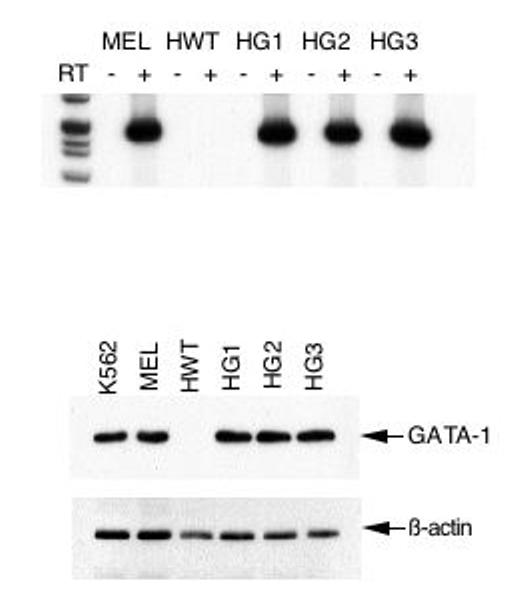
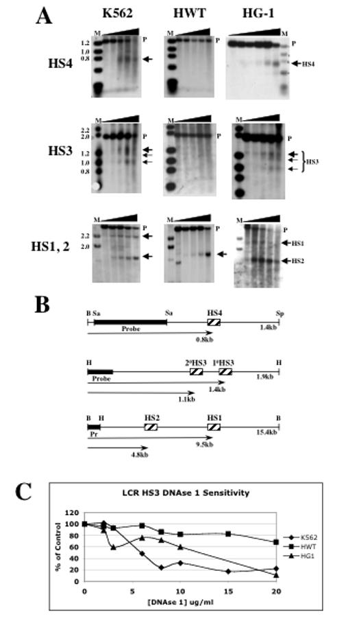
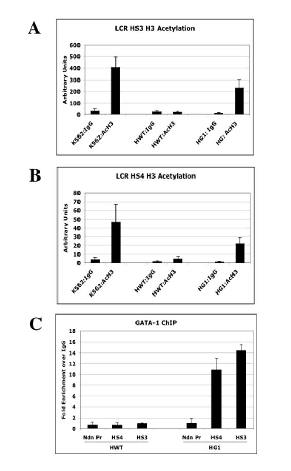
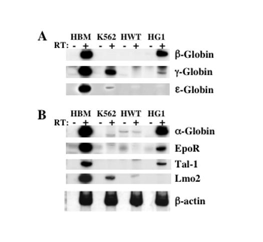
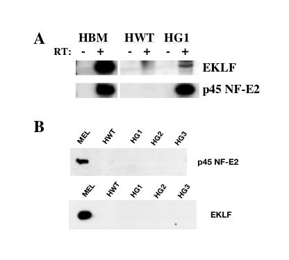
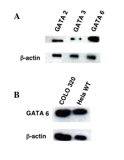
GATA-1 is a hematopoietic transcription factor expressed in erythroid, megakaryocytic, mast cell and eosinophil lineages. It is required for normal erythroid differentiation, the expression of erythroid-specific genes and for the establishment of an active chromatin structure throughout the beta-globin gene locus. GATA-1 is also necessary for the formation and function of the locus control region DNase I hypersensitive site (HS) core elements. To determine whether GATA-1 was sufficient to direct formation of the locus control region (LCR) and an erythroid pattern of gene expression, we expressed GATA-1 in the non-hematopoietic HeLa cell line that does not express other hematopoietic transcription factors but does express GATA-2, GATA-3, and GATA-6. We found that production of the GATA-1 protein resulted in the formation of LCR DNase I HSs 1-4 in their normal locations, and that histones became hyperacetylated within these regulatory elements. Transcription of several erythroid-specific genes was activated in HeLa cells expressing GATA-1, including those coding for alpha-globin, beta-globin, the erythropoietin receptor, the erythroid krüpple-like factor and p45 NF-E2. Despite increased expression of these genes at the mRNA level, their protein products were not detected. These results imply that GATA-1 is sufficient to direct chromatin structure reorganization within the beta-globin LCR and an erythroid pattern of gene expression in the absence of other hematopoietic transcription factors.
Figures
Similar articles
-
Beta-globin active chromatin Hub formation in differentiating erythroid cells and in p45 NF-E2 knock-out mice.J Biol Chem. 2007 Jun 1;282(22):16544-52. doi: 10.1074/jbc.M701159200. Epub 2007 Apr 11. J Biol Chem. 2007. PMID: 17428799
-
The distinctive roles of erythroid specific activator GATA-1 and NF-E2 in transcription of the human fetal γ-globin genes.Nucleic Acids Res. 2011 Sep 1;39(16):6944-55. doi: 10.1093/nar/gkr253. Epub 2011 May 24. Nucleic Acids Res. 2011. PMID: 21609963 Free PMC article.
-
Erythroid activator NF-E2, TAL1 and KLF1 play roles in forming the LCR HSs in the human adult β-globin locus.Int J Biochem Cell Biol. 2016 Jun;75:45-52. doi: 10.1016/j.biocel.2016.03.013. Epub 2016 Mar 26. Int J Biochem Cell Biol. 2016. PMID: 27026582
-
Erythroid regulatory elements.Stem Cells. 1993 Mar;11(2):95-104. doi: 10.1002/stem.5530110204. Stem Cells. 1993. PMID: 8096156 Review.
-
ChIPs of the beta-globin locus: unraveling gene regulation within an active domain.Curr Opin Genet Dev. 2002 Apr;12(2):170-7. doi: 10.1016/s0959-437x(02)00283-6. Curr Opin Genet Dev. 2002. PMID: 11893490 Review.
Cited by
-
Progress in detecting cell-surface protein receptors: the erythropoietin receptor example.Ann Hematol. 2014 Feb;93(2):181-92. doi: 10.1007/s00277-013-1947-2. Epub 2013 Dec 14. Ann Hematol. 2014. PMID: 24337485 Free PMC article. Review.
-
Nkx2-5 represses Gata1 gene expression and modulates the cellular fate of cardiac progenitors during embryogenesis.Circulation. 2011 Apr 19;123(15):1633-41. doi: 10.1161/CIRCULATIONAHA.110.008185. Epub 2011 Apr 4. Circulation. 2011. PMID: 21464046 Free PMC article.
-
Developmental- and differentiation-specific patterns of human gamma- and beta-globin promoter DNA methylation.Blood. 2007 Aug 15;110(4):1343-52. doi: 10.1182/blood-2007-01-068635. Epub 2007 Apr 24. Blood. 2007. PMID: 17456718 Free PMC article.
-
Epo receptors are not detectable in primary human tumor tissue samples.PLoS One. 2013 Jul 4;8(7):e68083. doi: 10.1371/journal.pone.0068083. Print 2013. PLoS One. 2013. PMID: 23861852 Free PMC article.
-
Hypoxia up-regulates expression of hemoglobin in alveolar epithelial cells.Am J Respir Cell Mol Biol. 2011 Apr;44(4):439-47. doi: 10.1165/rcmb.2009-0307OC. Epub 2010 May 27. Am J Respir Cell Mol Biol. 2011. PMID: 20508070 Free PMC article.
References
-
- Orkin SH. GATA-binding transcription factors in hematopoietic cells. Blood. 1992;80:575–81. - PubMed
-
- Cantor AB, Orkin SH. Transcriptional regulation of erythropoiesis: an affair involving multiple partners. Oncogene. 2002;21:3368–76. - PubMed
-
- Pevny L, Simon MC, Robertson E, Klein WH, Tsai SF, D'Agati V, Orkin SH, Costantini F. Erythroid differentiation in chimaeric mice blocked by a targeted mutation in the gene for transcription factor GATA-1. Nature. 1991;349:257–60. - PubMed
Publication types
MeSH terms
Substances
Grants and funding
LinkOut - more resources
Full Text Sources
Molecular Biology Databases
