. 2007 Jan 9;104(2):630-5.
doi: 10.1073/pnas.0608341104. Epub 2006 Dec 29.

par genes and the pathology of chromosome loss in Vibrio cholerae

Affiliations

par genes and the pathology of chromosome loss in Vibrio cholerae

Yoshiharu Yamaichi et al. Proc Natl Acad Sci U S A. .

Abstract

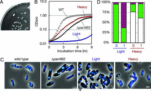
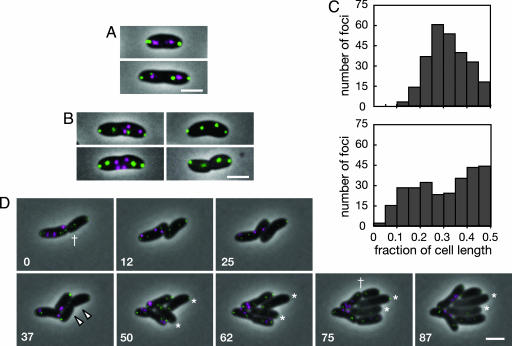
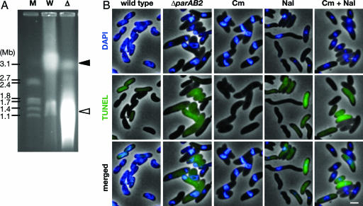
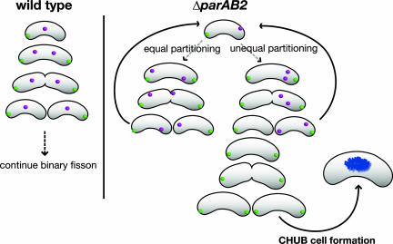
The causes and consequences of chromosome loss in bacteria with multiple chromosomes are unknown. Vibrio cholerae, the causative agent of the severe diarrheal disease cholera, has two circular chromosomes. Like many other bacterial chromosomes, both V. cholerae chromosomes contain homologues of plasmid partitioning (par) genes. In plasmids, par genes act to segregate plasmid molecules to daughter cells and thereby ensure plasmid maintenance; however, the contribution of par genes to chromosome segregation is not clear. Here, we show that the chromosome II parAB2 genes are essential for the segregation of chromosome II but not chromosome I. In a parAB2 deletion mutant, chromosome II is mislocalized and frequently fails to segregate, yielding cells with only chromosome I. These cells divide once; their progeny are not viable. Instead, chromosome II-deficient cells undergo dramatic cell enlargement, nucleoid condensation and degradation, and loss of membrane integrity. The highly consistent nature of these cytologic changes suggests that prokaryotes, like eukaryotes, may possess characteristic death pathways.


Conflict of interest statement

The authors declare no conflict of interest.

Figures

Fig. 1.
Fig. 1.
V. cholerae parABS2 stabilizes a mini-F plasmid in E. coli. Stability is measured as the percentage of cells harboring mini-F plasmid derivatives over time. The mini-F derivatives are pYB145 (parABS2+; filled squares, solid line), pYB095 (parAB2+; filled circles, gray line), pYB141 (parS2+; open squares, broken line), and control vector pXX705 (open circles, dotted line).
Fig. 2.
Fig. 2.
Growth defect and heterogeneity of cell size, shape, and nucleoid morphology in a ΔparAB2 V. cholerae strain. (A) Heterogeneous colony size of a ΔparAB2 strain harboring a parAB2 complementing plasmid grown on nonselective LB plates. All of the normal-sized colonies were found to carry the parAB2-bearing plasmid, whereas all of the tiny colonies (arrowhead) lost the parAB2-bearing plasmid (data not shown). (B) Growth in LB at 37°C of WT (diamonds), unfractionated ΔparAB2 cells (black circles), light fraction (blue circles), and heavy fraction (red circles). (C) Merged phase-contrast and fluorescence images of DAPI-stained fixed cells. The arrowheads point to cells with fragmented nucleoids. (Scale bar, 2 μm.) (D) BacLight live/dead assay of fractionated cells. Cells were assayed immediately after fractionation (time 0) or after growth in LB medium at 37°C for 1 h. Normal and CHUB cells were distinguished by morphology. Depicted are normal-live cells (white), normal-dead cells (black), CHUB-live cells (green), and CHUB-dead cells (magenta).
Fig. 3.
Fig. 3.
Number and subcellular location of YFP-ParB1 and CFP-ParB2 foci in WT and ΔparAB2 V. cholerae. (A and B) Static images of representative WT (A) and ΔparAB2 (B) cells expressing YFP-ParB1 and CFP-ParB2. (C) Histograms showing the localization of CFP-ParB2 foci in WT (Upper) and ΔparAB2 (Lower) cells. The distance from each focus to the nearest pole was measured and divided by cell length. A total of 261 foci from 118 WT cells and 274 foci from 103 ΔparAB2 cells were analyzed. (D) Time-lapse images of ΔparAB2 cells taken at 12-min intervals at 35°C. The number in the lower left corner of each image indicates the time (min). YFP is shown in green, and CFP is shown in magenta. The dagger shows a cell division yielding a cell lacking chrII. The arrowheads show cells that will become CHUBs. Asterisks show enlarging CHUB cells. (Scale bar, 2 μm.)
Fig. 4.
Fig. 4.
ChrI degradation in CHUB cells. (A) Pulsed-field gel electrophoresis of intact (undigested) genomic DNA prepared from WT (W) and ΔparAB2 (Δ) cells. Molecular weight standards (M) are shown at left. The positions of chrI and chrII are indicated by the black and white arrowheads, respectively. In principal, the more rapidly migrating DNA in the ΔparAB2 cells could be derived from increased initiations of replication of chrI; however, overinitiation of chrI does not appear to occur in ΔparAB2 cells, inasmuch as the ratio oriCIvc/TerIvc in these cells was 0.88, compared with 1.0 in WT, as determined by qPCR. (B) TUNEL assay of ΔparAB2 cells and WT cells with no treatment or with treatment with chloramphenicol (Cm), nalidixic acid (Nal), or both. (Scale bar, 2 μm.)
Fig. 5.
Fig. 5.
Schematic representation of chromosome segregation and the generation of CHUB cells in ΔparAB2 V. cholerae. (Left) Schematic representation showing the pattern of segregation of the origin regions of chrI (green) and chrII (magenta) in WT cells. (Right) The chrII origin region is randomly localized in the ΔparAB2 mutant. Cell division can therefore result in daughter cells that either contain or lack this chromosome. Cells lacking chrII divide only once, and then their progeny develop into CHUB cells.

References

    1. Ogura T, Hiraga S. Cell. 1983;32:351–360. - PubMed
    1. Austin S, Abeles A. J Mol Biol. 1983;169:373–387. - PubMed
    1. Niki H, Hiraga S. Cell. 1997;90:951–957. - PubMed
    1. Li Y, Dabrazhynetskaya A, Youngren B, Austin S. Mol Microbiol. 2004;53:93–102. - PubMed
    1. Ebersbach G, Gerdes K. Annu Rev Genet. 2005;39:453–479. - PubMed

Publication types

Substances

LinkOut - more resources