. 2007 Jan;48(1):446-54.
doi: 10.1167/iovs.06-0190.

Retinal stem cells transplanted into models of late stages of retinitis pigmentosa preferentially adopt a glial or a retinal ganglion cell fate

Affiliations

Retinal stem cells transplanted into models of late stages of retinitis pigmentosa preferentially adopt a glial or a retinal ganglion cell fate

Kriss Canola et al. Invest Ophthalmol Vis Sci. 2007 Jan.

Abstract

Purpose: To characterize the potential of newborn retinal stem cells (RSCs) isolated from the radial glia population to integrate the retina, this study was conducted to investigate the fate of in vitro expanded RSCs transplanted into retinas devoid of photoreceptors (adult rd1 and old VPP mice and rhodopsin-mutated transgenic mice) or partially degenerated retina (adult VPP mice) retinas.

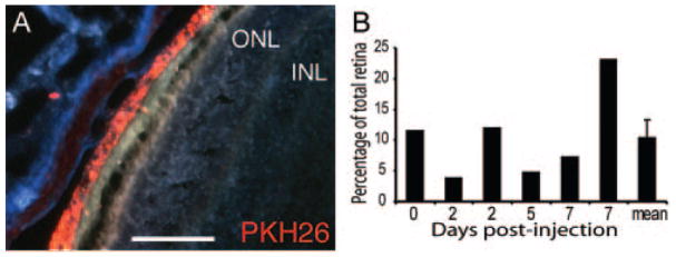
Methods: Populations of RSCs and progenitor cells were isolated either from DBA2J newborn mice and labeled with the red lipophilic fluorescent dye (PKH26) or from GFP (green fluorescent protein) transgenic mice. After expansion in EGF+FGF2 (epidermal growth factor+fibroblast growth factor), cells were transplanted intravitreally or subretinally into the eyes of adult wild-type, transgenic mice undergoing slow (VPP strain) or rapid (rd1 strain) retinal degeneration.

Results: Only limited migration and differentiation of the cells were observed in normal mice injected subretinally or in VPP and rd1 mice injected intravitreally. After subretinal injection in old VPP mice, transplanted cells massively migrated into the ganglion cell layer and, at 1 and 4 weeks after injection, harbored neuronal and glial markers expressed locally, such as beta-tubulin-III, NeuN, Brn3b, or glial fibrillary acidic protein (GFAP), with a marked preference for the glial phenotype. In adult VPP retinas, the grafted cells behaved similarly. Few grafted cells stayed in the degenerating outer nuclear layer (ONL). These cells were, in rare cases, positive for rhodopsin or recoverin, markers specific for photoreceptors and some bipolar cells.

Conclusions: These results show that the grafted cells preferentially integrate into the GCL and IPL and express ganglion cell or glial markers, thus exhibiting migratory and differentiation preferences when injected subretinally. It also appears that the retina, whether partially degenerated or already degenerated, does not provide signals to induce massive differentiation of RSCs into photoreceptors. This observation suggests that a predifferentiation of RSCs into photoreceptors before transplantation may be necessary to obtain graft integration in the ONL.


Figures

Figure 1
Figure 1
Cell localization after transplantation in retinas devoid of photoreceptors showed only slight migration when injected intravitreally in VPP as well as in rd1 mice. (A) By 1 week after injection, RSCs intravitreally injected into the VPP mouse retina formed elongated clusters located in close proximity to the GCL but not within. (B) By 4 weeks after surgical intervention, some grafted cells had migrated within the GCL. In the rd1 mice, transplanted cells showed similar localization equally close to the GCL during the week after injection (C) and slight migration within it 4 weeks after transplantation (D). Moreover, intravitreal injection allowed the cells to migrate into various structures of the rd1 eye such as the iris (E) and the CB (F), which is not the case with subretinal injections (not shown).
Figure 2
Figure 2
Assessment of the subretinal injection in the normal retina. The size of the graft is expressed as a percentage of the total surface of the retina in six retinas from 0 to 7 days after injection (A). Subretinal injection of RSCs into the eye of C57Bl/6 mice shows that the cells labeled with the PKH26 were restricted to the SS and were not scattered in other layers of the retina 2 days after the surgery (B). Scale bar, 50 μm.
Figure 3
Figure 3
RSCs massively migrated toward the innermost layer of the retina after subretinal injection into fully degenerated VPP retinas. Compared with the poor migration of intravitreally injected cells (Fig. 1), subretinal injections exhibited a massive migration of the RSCs toward the GCL of VPP retinas. This migration was already observable by 1 week after surgery, with cells present in the subretinal space (the site of injection) as well as in all the other layers of the retina up to the GCL with GFP-expressing RSCs (A) and PKH26-labeled RSCs (B). The migration into the GCL was completed by 4 weeks after injection (C). Scale bars: (A, B) 50 μm; (C) 100 μm.
Figure 4
Figure 4
RSCs expressed neuronal and neuroretinal markers after transplantation in a fully degenerated retina. (A1, B1, C1, F1) Fluorescence images of degenerated retinal sections showing DAPI staining (blue) of the nuclei and PKH26-labeled RSCs (red) within the host GCL; (A2, B2, C2, F2) grafted cells and host retina labeled with specific antibodies revealed by secondary antibodies coupled to FITC (green) and nuclei labeled with DAPI; (A3, B3, C3, F3) merged images showing colocalization of the PKH26 (red) with the antibody labeling (green) and the DAPI staining (blue). RSCs injected subretinally incorporated into the GCL and expressed the early neuronal marker β-tubulin-III (A). The RSCs that incorporated into the GCL expressed Brn3b (B) a marker for mature ganglion cells (inset: positive control for Brn3b labeling, note that Brn3b labels a subpopulation of RGC). Integrated RSCs also express NeuN (C) a specific marker for ganglion cell lineage in the retina as shown in confocal images (C). Colocalization of these antibodies (Brn3b and NeuN) and the PKH26 among the host retina was observed exclusively in the GCL, denoting a region of specific immunoreactivity. Insets: higher magnification of the area defined by the arrows (D, E). Fluorescence images of GFP-expressing RSCs subretinally injected and exhibiting morphologies resembling horizontal cells (D1, D2, arrowheads) close to calbindin-positive cells (a specific marker for horizontal cells, D2, red, arrow) but negative for the latter, even if exhibiting a similar morphology, and with bipolar cells (E, arrow) correctly located and oriented in the INL. Occasionally, RSCs that did not migrate toward the GCL but stayed close to the INL, where few remaining photoreceptors were located, expressed rhodopsin, a specific marker for photoreceptors (F, confocal image). Scale bar: (A, B) 25 μm; (C, F) 30 μm; (D) 10 μm; (E) 100 μm.
Figure 5
Figure 5
Glial cell differentiation after transplantation. (A1) Fluorescence images of retinal sections showing PKH26-labeled RSCs (red) within the host degenerated retina and DAPI staining (blue) of the nuclei; (A2) grafted cells and host retina labeled with anti-GFAP revealed by a secondary antibody coupled to FITC (green) and nuclei labeled with DAPI; (A3) merged image showing colocalization of the PKH26 (red) with the antibody labeling (green) and the DAPI staining (blue). Colocalization depicts a grafted cell positive for the glial marker GFAP within the host GCL after subretinal injection (A). The preference for the glial phenotype is supported by the colocalization of PKH26 and GFAP in cells integrated after intravitreal injections. This preference is further supported by the high degree of GFAP expression (green) by grafted (red) cells that have migrated close to the lens (B) after intravitreal delivery of RSCs. Endogenous glia revealed by GFAP immunostaining (green) in a control retina, labels only end feet of Müller cells and the astrocytes contained in the GCL (C). In comparison, in transplanted retina, 4 weeks after surgery, extensive gliosis related to increased GFAP labeling (green) is observed (D) in the site of injection (arrowheads) up to the site where grafted cells (red) incorporate (). DAPI staining to visualize nuclei was performed on each section (blue). Scale bar, (A) 25 μm; (B) 50 μm; (C, D) 100 μm.
Figure 6
Figure 6
RSCs transplanted to partially degenerated retina (7-month-old VPP) did not integrate better into the ONL and showed a higher tendency to acquire glial characteristics than cells transplanted into fully degenerated retina. The ONL of 7-month retinas presented one to six rows of photoreceptors, illustrating the heterogeneity of the degeneration as shown in retinal sections from the same retinal area of two animals of the same age (A1, A2). Intravitreal injection in the partially degenerated retina was rigorously similar to the fully degenerated retina by 1 week and 4 weeks after injection (B, C, respectively). After subretinal injections, grafted RSCs had massively migrated toward the GCL and expressed the glial marker, GFAP (D1, D2, D3). Indeed, GFP expressing RSCs (green, D1) and GFAP immunostaining (red, D2) colocalized, shown in a merged image (orange, D3). Thus, RSCs transplanted into the partially degenerated 7-month-old VPP mouse retina behaved almost like those in their counterparts at 10 months of age. Nevertheless, 4 weeks after transplantation, some subretinally injected RSCs stayed in the remaining ONL (E1, arrowhead) and in rare cases express the recoverin marker (E2, E3, arrowhead). DAPI staining to visualize nuclei was performed on each section. Scale bar: (A) 25 μm; (BE) 50 μm.

References

    1. Rivolta C, Sharon D, DeAngelis MM, Dryja TP. Retinitis pigmentosa and allied diseases: numerous diseases, genes, and inheritance patterns. Hum Mol Genet. 2002;11:1219–1227. - PubMed
    1. Delyfer MN, Leveillard T, Mohand-Said S, Hicks D, Picaud S, Sahel JA. Inherited retinal degenerations: therapeutic prospects. Biol Cell. 2004;96:261–269. - PubMed
    1. Nishida A, Takahashi M, Tanihara H, et al. Incorporation and differentiation of hippocampus-derived neural stem cells transplanted in injured adult rat retina. Invest Ophthalmol Vis Sci. 2000;41:4268–4274. - PubMed
    1. Takahashi M, Palmer TD, Takahashi J, Gage FH. Widespread integration and survival of adult-derived neural progenitor cells in the developing optic retina. Mol Cell Neurosci. 1998;12:340–348. - PubMed
    1. Young MJ, Ray J, Whiteley SJ, Klassen H, Gage FH. Neuronal differentiation and morphological integration of hippocampal progenitor cells transplanted to the retina of immature and mature dystrophic rats. Mol Cell Neurosci. 2000;16:197–205. - PubMed

Publication types

MeSH terms