

Severe steatosis increases hepatocellular injury and impairs liver regeneration in a rat model of partial hepatectomy
- PMID: 17197964
- PMCID: PMC1867939
- DOI: 10.1097/01.sla.0000225253.84501.0e
Severe steatosis increases hepatocellular injury and impairs liver regeneration in a rat model of partial hepatectomy
Abstract
Objective: The aim of this study was to assess the influence of severe steatosis with inflammation on hepatocellular recovery after 70% hepatectomy in a rat model of diet-induced steatosis.
Background: Patients with steatosis have an increased risk of inflammatory complications after liver resection. This might be attributable to Kupffer cell-mediated inflammation in steatotic livers causing progressive injury.
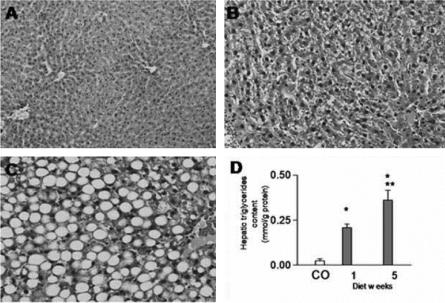
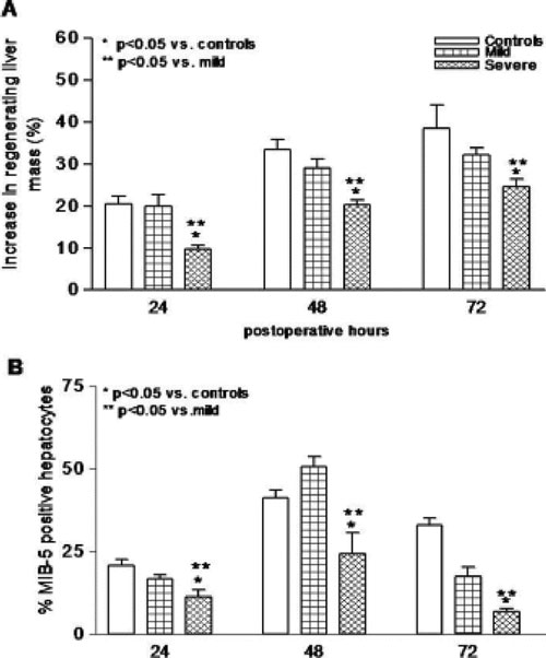
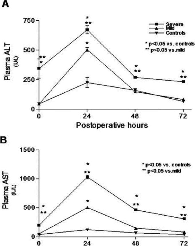
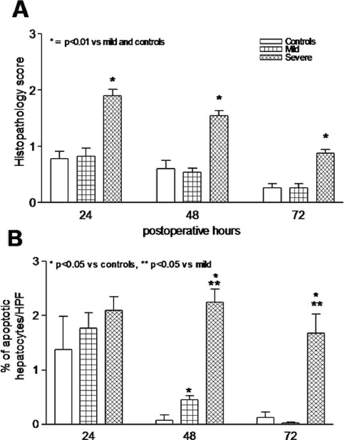
Methods: Male Wistar rats were fed a standard methionine- and choline-deficient diet for 1 or 5 weeks. A 70% partial hepatectomy (PH) was performed, after which rats were killed at 24, 48, or 72 hours. The extent of steatosis and inflammation was determined by assessment of hepatic triglycerides, cytokine content, and histopathology. Outcome parameters were: liver regeneration (MIB-5 proliferation rate, mitotic index, and regenerating liver mass), hepatocellular injury (plasma aminotransferases, lipid peroxidation, histopathology, and apoptosis), Kupffer cell-mediated proinflammatory response (TNF-alpha, IL-1beta, IL-6, IL-10 in plasma and liver) and antioxidant content (total glutathione).
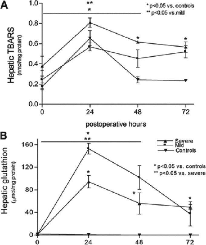
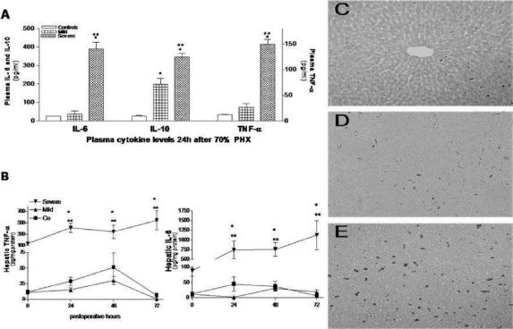
Results: Methionine- and choline-deficient diet induced uncomplicated steatosis after 1 week (<30% hepatocytes affected without inflammation) and severe steatosis after 5 weeks (>60% hepatocytes affected, including prominent inflammation) as confirmed by histopathology. After PH, liver regeneration was impaired at all time points in the severe steatosis group as compared with the mild and control groups (P < 0.05). Hepatocellular injury was significantly increased in the severe steatosis group at all time points (P < 0.05). Kupffer cell-mediated inflammatory responses were aggravated in the severe steatosis group along with decreased antioxidant content (P < 0.05). Necrosis was the main type of cell death in severe steatotic livers compared with mainly apoptotic cell death in mild steatotic and normal livers.
Conclusion: Steatosis with prominent inflammation impaired liver regeneration probably because of increased hepatocellular lipid peroxidation and damage in concert with Kupffer cell-mediated proinflammatory responses. These results suggest an increased risk of performing extensive liver resection in the presence of severe steatosis.
Figures
Similar articles
-
Hepatic regeneration and functional recovery following partial liver resection in an experimental model of hepatic steatosis treated with omega-3 fatty acids.Br J Surg. 2013 Apr;100(5):674-83. doi: 10.1002/bjs.9059. Br J Surg. 2013. PMID: 23456631
-
Mild steatosis impairs functional recovery after liver resection in an experimental model.Br J Surg. 2007 Aug;94(8):1002-8. doi: 10.1002/bjs.5672. Br J Surg. 2007. PMID: 17497653
-
Impaired liver regeneration of steatotic rats after portal vein ligation: a particular emphasis on (99m)Tc-DISIDA scintigraphy and adiponectin signaling.J Hepatol. 2010 Apr;52(4):540-9. doi: 10.1016/j.jhep.2010.01.005. Epub 2010 Feb 19. J Hepatol. 2010. PMID: 20206399
-
Fatty liver in liver transplantation and surgery.Semin Liver Dis. 2001;21(1):105-13. doi: 10.1055/s-2001-12933. Semin Liver Dis. 2001. PMID: 11296690 Review.
-
The role of the mTOR pathway during liver regeneration and tumorigenesis.Ann Endocrinol (Paris). 2013 May;74(2):121-2. doi: 10.1016/j.ando.2013.03.003. Epub 2013 Apr 6. Ann Endocrinol (Paris). 2013. PMID: 23566619 Review.
Cited by
-
Leptin increases mitotic index and regeneration ratio in hepatectomized rats.Med Sci Monit Basic Res. 2013 Nov 13;19:279-84. doi: 10.12659/MSMBR.889591. Med Sci Monit Basic Res. 2013. PMID: 24220642 Free PMC article.
-
Loss of L-selectin-guided CD8+ , but not CD4+ , cells protects against ischemia reperfusion injury in a steatotic liver.Hepatology. 2017 Oct;66(4):1258-1274. doi: 10.1002/hep.29276. Epub 2017 Sep 4. Hepatology. 2017. PMID: 28543181 Free PMC article.
-
Simultaneous assessment of liver volume and whole liver fat content: a step towards one-stop shop preoperative MRI protocol.Eur Radiol. 2011 Feb;21(2):301-9. doi: 10.1007/s00330-010-1941-1. Epub 2010 Sep 3. Eur Radiol. 2011. PMID: 20814683
-
Administration of multipotent mesenchymal stromal cells restores liver regeneration and improves liver function in obese mice with hepatic steatosis after partial hepatectomy.Stem Cell Res Ther. 2017 Jan 28;8(1):20. doi: 10.1186/s13287-016-0469-y. Stem Cell Res Ther. 2017. PMID: 28129776 Free PMC article.
-
The effect of preoperative chemotherapy on liver regeneration after portal vein embolization/ligation or liver resection in patients with colorectal liver metastasis: a systematic review protocol.Syst Rev. 2020 Dec 4;9(1):279. doi: 10.1186/s13643-020-01545-w. Syst Rev. 2020. PMID: 33276812 Free PMC article.
References
-
- Scheele J, Stangl R, Altendorf-Hofmann A. Hepatic metastases from colorectal carcinoma: impact of surgical resection on the natural history. Br J Surg. 1990;77:1241–1246. - PubMed
-
- Paquet KJ, Koussouris P, Mercado MA, et al. Limited hepatic resection for selected cirrhotic patients with hepatocellular or cholangiocellular carcinoma: a prospective study. Br J Surg. 1991;78459–78462. - PubMed
-
- Wu CC, Yang MD, Liu TJ. Improvements in hepatocellular carcinoma resection by intraoperative ultrasonography or intermittent hepatic inflow blood occlusion. Jpn Clin Oncol. 1992;22:107–111. - PubMed
-
- Hilden M, Christofferson P, Juhl E. Liver histology in a ‘normal population’: examination of 503 consecutive fatal traffic casualties. Scand J Gastroenterol. 1977;12:593–597. - PubMed
-
- Neuschwander-Tetri BA, Caldwell SH. Nonalcoholic steatohepatitis: summary of an AASLD Single Topic Conference. Hepatology. 2003;37:1202–1219. - PubMed
MeSH terms
LinkOut - more resources
Full Text Sources
