Successful iliac vein and inferior vena cava stenting ameliorates venous claudication and improves venous outflow, calf muscle pump function, and clinical status in post-thrombotic syndrome
- PMID: 17197976
- PMCID: PMC1867924
- DOI: 10.1097/01.sla.0000245550.36159.93
Successful iliac vein and inferior vena cava stenting ameliorates venous claudication and improves venous outflow, calf muscle pump function, and clinical status in post-thrombotic syndrome
Abstract
Objectives: Stent therapy has been proposed as an effective treatment of chronic iliofemoral (I-F) and inferior vena cava (IVC) thrombosis. The purpose of this study was to determine the effects of technically successful stenting in consecutive patients with advanced CVD (CEAP3-6 +/- venous claudication) for chronic obliteration of the I-F (+/-IVC) trunks, on the venous hemodynamics of the limb, the walking capacity, and the clinical status of CVD. These patients had previously failed to improve with conservative treatment entailing compression and/or wound care for at least 12 months.
Methods: The presence of venous claudication was assessed by > or =3 independent examiners. The CEAP clinical classification was used to determine the severity of CVD. Outflow obstruction [Outflow Fraction at 1- and 4-second (OF1 and OF4) in %], venous reflux [Venous Filling Index (VFI) in mL/100 mL/s], calf muscle pump function [Ejection Fraction (EF) in %] and hypertension [Residual Venous Fraction (RVF) in %], were examined before and after successful venous stenting in 16 patients (23 limbs), 6 females, 10 males, median age 42 years; range, 31-77 yearas, left/right limbs 14/9, using strain gauge plethysmography; 7/16 of these had thrombosis extending to the IVC. Contralateral limbs to those stented without prior I-F +/- IVC thrombosis, nor infrainguinal clots on duplex, were used as control limbs (n = 9). Excluded were patients with stent occlusion or stenoses, peripheral arterial disease (ABI <1.0), symptomatic cardiac disease, unrelated causes of walking impairment, and malignancy. Preinterventional data (< or =30 days) were compared with those after endovascular therapy (8.4 months; interquartile range [IQR], 3-11.8 months). Nonparametric analysis was applied.
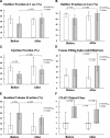
Results: Compared with the control group, limbs with I-F +/- IVC thrombosis before stenting had reduced venous outflow (OF4) and calf muscle pump function (EF), worse CEAP clinical class, and increased RVF (all, P < 0.05). At 8.4 months (IQR, 3-11.8 months) after successful I-F (+/-IVC) stenting, venous outflow (OF1, OF4) and calf muscle pump function (EF) had both improved (P < 0.001) and the RVF had decreased (P < 0.001), at the expense of venous reflux, which had increased further (increase of median VFI by 24%; P = 0.002); the CEAP status had also improved (P < 0.05) from a median class C3 (range, C3-C6; IQR, C3-C5) [distribution, C6: 6; C4: 4; C3: 13] before intervention to C2 (range, C2-C6; IQR, C2-C4.5) [distribution, C6: 1; C5: 5; C4: 4; C2: 13] after intervention. At this follow up (8.4 months median), venous outflow (OF1, OF4), calf muscle pump function (EF), and RVF of the stented limbs did not differ significantly from those of the control; significantly worse (P < 0.025) were the amount of venous reflux (VFI), and the CEAP clinical class, despite the improvement with stenting. Incapacitating venous claudication noted in 62.5% (10 of 16, 95% CI, 35.8%-89.1%) of patients (15 of 23 limbs; 65.2%, 95% CI, 44.2%-86.3%) before stenting was eliminated in all after stenting (P < 0.001).
Conclusions: Successful I-F (+/-IVC) stenting in limbs with venous outflow obstruction and complicated CVD (C3-C6) ameliorates venous claudication, normalizes outflow, and enhances calf muscle pump function, compounded by a significant clinical improvement of CVD. The significant increase in the amount of venous reflux of the stented limbs indicates that elastic or inelastic compression support of the successfully stented limbs would be pivotal in preventing disease progression.
Figures


Similar articles
-
Hemodynamic impairment, venous segmental disease, and clinical severity scoring in limbs with Klippel-Trenaunay syndrome.J Vasc Surg. 2007 Mar;45(3):561-7. doi: 10.1016/j.jvs.2006.11.032. Epub 2007 Jan 31. J Vasc Surg. 2007. PMID: 17275246
-
Iliac-femoral venous stenting for lower extremity venous stasis symptoms.Ann Vasc Surg. 2012 Feb;26(2):185-9. doi: 10.1016/j.avsg.2011.05.033. Epub 2011 Oct 22. Ann Vasc Surg. 2012. PMID: 22018502
-
Contemporary outcomes of elective iliocaval and infrainguinal venous intervention for post-thrombotic chronic venous occlusive disease.J Vasc Surg Venous Lymphat Disord. 2017 Nov;5(6):789-799. doi: 10.1016/j.jvsv.2017.05.020. J Vasc Surg Venous Lymphat Disord. 2017. PMID: 29037346
-
Treatment of iliac-caval outflow obstruction.Semin Vasc Surg. 2015 Mar;28(1):47-53. doi: 10.1053/j.semvascsurg.2015.07.001. Epub 2015 Jul 17. Semin Vasc Surg. 2015. PMID: 26358309 Review.
-
Endovascular management of venous ulcer in a patient with occluded duplicated inferior vena cava and review of inferior vena cava development.Vasc Endovascular Surg. 2014 Feb;48(2):162-5. doi: 10.1177/1538574413510627. Epub 2013 Nov 12. Vasc Endovascular Surg. 2014. PMID: 24226789 Review.
Cited by
-
Iliac vein compression: epidemiology, diagnosis and treatment.Vasc Health Risk Manag. 2019 May 9;15:115-122. doi: 10.2147/VHRM.S203349. eCollection 2019. Vasc Health Risk Manag. 2019. PMID: 31190849 Free PMC article. Review.
-
Dramatic recovery of chronic non-healing ulcer secondary to recurrent unprovoked DVT by venous stenting.BMJ Case Rep. 2014 May 14;2014:bcr2014204145. doi: 10.1136/bcr-2014-204145. BMJ Case Rep. 2014. PMID: 24827830 Free PMC article. No abstract available.
-
Unraveling the pathophysiology of lower-limb postthrombotic syndrome in adolescents: a proof-of-concept study.Blood Adv. 2023 Jun 27;7(12):2784-2793. doi: 10.1182/bloodadvances.2022009599. Blood Adv. 2023. PMID: 36763520 Free PMC article.
-
Safety and Feasibility of the RevCore Catheter for Venous In-Stent Thrombosis: A Multicenter, Retrospective Study.J Soc Cardiovasc Angiogr Interv. 2025 Mar 20;4(4):102571. doi: 10.1016/j.jscai.2025.102571. eCollection 2025 Apr. J Soc Cardiovasc Angiogr Interv. 2025. PMID: 40308240 Free PMC article.
-
Endovascular stent placement for chronic post-thrombotic symptomatic ilio-femoral venous obstructive lesions: a single-center study of safety, efficacy and quality-of-life improvement.Quant Imaging Med Surg. 2016 Aug;6(4):342-352. doi: 10.21037/qims.2016.07.07. Quant Imaging Med Surg. 2016. PMID: 27709070 Free PMC article.
References
-
- Akesson H, Brudin L, Dahlstrom JA, et al. Venous function assessed during a 5 year period after iliofemoral venous thrombosis treated with anticoagulation. Eur J Vasc Surg. 1990;4:43–48. - PubMed
-
- Adams JG, Silver D. Deep venous thrombosis and pulmonary embolism. In: Dean RH, Yao JST, Brewster DC, eds. Current Diagnosis and Treatment in Vascular Surgery. London: Prentice-Hall, 1995:375–390.
-
- Comerota AJ, Throm RC, Mathias SD, et al. Catheter-directed thrombolysis for iliofemoral deep venous thrombosis improves health-related quality of life. J Vasc Surg. 2000;32:130–137. - PubMed
-
- Comerota AJ. Quality-of-life improvement using thrombolytic therapy for iliofemoral deep venous thrombosis. Rev Cardiovasc Med. 2002;3(suppl 2):61–67. - PubMed
MeSH terms
LinkOut - more resources
Full Text Sources
Other Literature Sources
Medical
Miscellaneous

