Review
. 2007 Jan 9;46(1):1-10.
doi: 10.1021/bi0620959.

Plasticity of amyloid fibrils

Affiliations
Review

Plasticity of amyloid fibrils

Ronald Wetzel et al. Biochemistry. .

Abstract

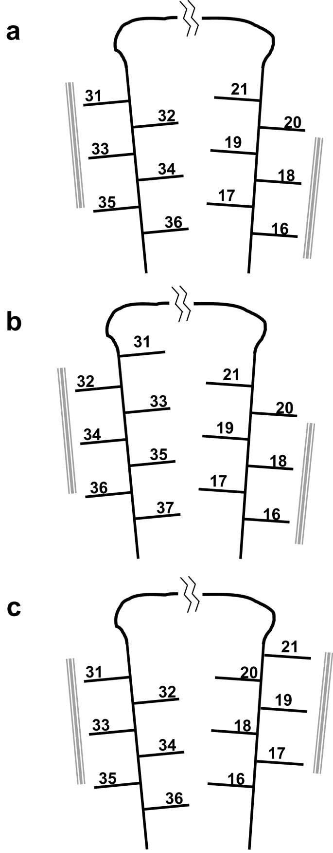
In experiments designed to characterize the basis of amyloid fibril stability through mutational analysis of the Abeta (1-40) molecule, fibrils exhibit consistent, significant structural malleability. In these results, and in other properties, amyloid fibrils appear to more resemble plastic materials generated from synthetic polymers than globular proteins. Thus, like synthetic polymers and plastics, amyloid fibrils exhibit both polymorphism, the ability of one polypeptide to form aggregates of different morphologies, and isomorphism, the ability of different polypeptides to grow into a fibrillar amyloid morphology. This view links amyloid with the prehistorical and 20th century use of proteins as starting materials to make films, fibers, and plastics, and with the classic protein fiber stretching experiments of the Astbury group. Viewing amyloids from the point of view of the polymer chemist may shed new light on a number of issues, such as the role of protofibrils in the mechanism of amyloid formation, the biological potency of fibrils, and the prospects for discovering inhibitors of amyloid fibril formation.


Figures

Figure 1
Figure 1
Elongation thermodynamics of the Aβ(1-40) amyloid fibril in response to alanine replacements. A. The amino acid sequence of Aβ(1-40). B. ΔΔGAla-WT of the elongation equilibrium for Ala mutants of Aβ(1-40). A positive value indicated destabilization of the mutant compared with wild type. The x-axis shows the numbered residue replaced with Ala. The * at position 23 indicates that this mutant peptide could not be dissolved and hence fibrils could not be grown under defined conditions. Adapted from reference (5), with inclusion of additional data (ADW and RW, previously unpublished) obtained using published methods (61).
Figure 2
Figure 2
(a) A model of a section of the Aβ(1-40) protofibril based on solid state NMR studies, based on pdb coordinates provided by Robert Tycko (7). (b) The G(β1) protein indicating residues 6 and 53 at adjacent positions in parallel β-sheet, from the pdb coordinates 2GB1. Figures rendered in PyMol (DeLano Scientific LLC).
Figure 3
Figure 3
Schematic renderings of different models for the orientation of side chains within the Aβ hairpin in amyloid fibrils. The numbered lines represent the side chains of amino acids involved in β-sheet within the fibril. The gray bars represent apparent intermolecular packing residues. Part a is reproduced from reference (13).
Figure 4
Figure 4
Elongation thermodynamics of multiple mutants of Aβ(1-40). (a) Additivity experiments of Ala mutants (see text). (b-d) Disulfide crosslinking experiments comparing the elongation equilibria of reduced (cross-hatched bars) and crosslinked, oxidized (open bars) double Cys mutants of Aβ(1-40) at the positions indicated. Part a is reproduced from reference (5) and part b from reference (13). Parts c and d based on previously unpublished data (SS and RW) obtained using published methods (62). Bars labeled “x + y” are mathematical sums of x and y data; bars labeled “x/y” show experimental data on x,y double mutants.
Figure 5
Figure 5
Proline mutagenesis of Aβ(1-40) amyloid structure and stability. (a) A plot of ΔΔGPro-WT with respect to sequence position of proline mutation. (b) A plot of ΔΔGPro-Ala with respect to sequence position. Diamonds indicate positions not analyzed; * indicates not measurable because of solubility problems. (a) and (b) reprinted from reference (5). (c) Hydrogen-exchange protection of amyloid fibrils from Pro mutants of Aβ(1-40). Higher deuterium exchange (y-axis) indicates less protection and therefore fewer backbone amide hydrogens in β-sheet in the fibrils. Reprinted from reference (4).
Figure 6
Figure 6
Schematic of the packing of interior residues in Aβ(1-40) fibrils from two different orientations of side chains within the Aβ hairpin. Parts A and D represent wild type Aβ (part a of Figure 3) and parts B and E represent an alternative conformation (part b of Fig. 3) in which the C-terminal extended chain (residues 31-36) is rotated 180°. Part C shows the basic parallel, in-register structure of the Aβ fibril. Green shaded residues indicated residues 16-21; tan/orange residues, residues 31-36. There is no side chain sphere for residue 33, which is Gly. Figure constructed by Sarina Bromberg.
Figure 7
Figure 7
Polymer spherulites. “Maltese cross” images in light microscopy. (a) Brain of a mouse model of human Aβ amyloidosis stained with Congo red and visualized by polarizing light microscopy (36); (b) Photomicrograph of a deformed isotactic polypropylene spherulite (courtesy R. Samuels). Reprinted from reference (63). (c) Environmental scanning electron microscopy image of insulin amyloid spherulites; scalebar 30 μm. From reference (64). (d) Phase microscope image of an isotactic polypropylene spherulite (courtesy R. Samuels). Reprinted from reference (63).

References

    1. Martin JB. Molecular basis of the neurodegenerative disorders [published erratum appears in N Engl J Med 1999 Oct 28;341(18):1407] N Engl J Med. 1999;340:1970–1980.
    1. Merlini G, Bellotti V. Molecular mechanisms of amyloidosis. N Engl J Med. 2003;349:583–596. - PubMed
    1. Wetzel R. Mutations and off-pathway aggregation. Trends in Biotechnology. 1994;12:193–198. - PubMed
    1. Williams AD, Portelius E, Kheterpal I, Guo JT, Cook KD, Xu Y, Wetzel R. Mapping abeta amyloid fibril secondary structure using scanning proline mutagenesis. J Mol Biol. 2004;335:833–842. - PubMed
    1. Williams AD, Shivaprasad S, Wetzel R. Alanine scanning mutagenesis of Abeta(1-40) amyloid fibril stability. J Mol Biol. 2006;357:1283–1294. - PubMed

Publication types