. 2007 Jan;12(1):99-112.
doi: 10.1016/j.devcel.2006.12.006.

Abnormal hair development and apparent follicular transformation to mammary gland in the absence of hedgehog signaling

Affiliations

Abnormal hair development and apparent follicular transformation to mammary gland in the absence of hedgehog signaling

Amel Gritli-Linde et al. Dev Cell. 2007 Jan.

Abstract

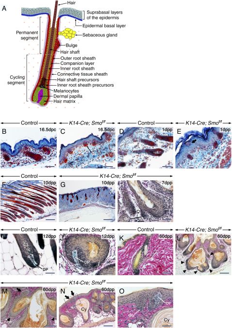
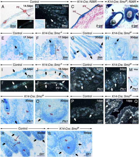
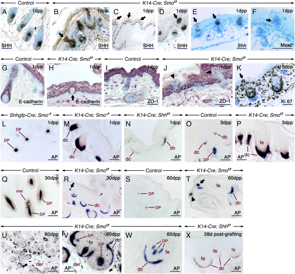
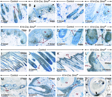
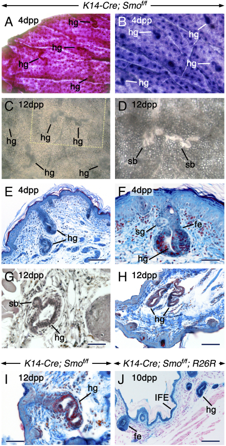
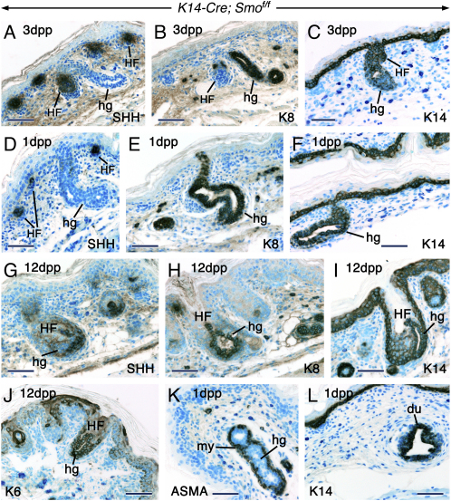
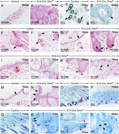
We show that removing the Shh signal tranducer Smoothened from skin epithelium secondarily results in excess Shh levels in the mesenchyme. Moreover, the phenotypes we observe reflect decreased epithelial Shh signaling, yet increased mesenchymal Shh signaling. For example, the latter contributes to exuberant hair follicle (HF) induction, while the former depletes the resulting follicular stem cell niches. This disruption of the niche apparently also allows the remaining stem cells to initiate hair formation at inappropriate times. Thus, the temporal structure of the hair cycle may depend on the physical structure of the niche. Finally, we find that the ablation of epithelial Shh signaling results in unexpected transformations: the follicular outer root sheath takes on an epidermal character, and certain HFs disappear altogether, having adopted a strikingly mammary gland-like fate. Overall, our study uncovers a multifaceted function for Shh in sculpting and maintaining the integrity and identity of the developing HF.


Figures

Figure 1
Figure 1
Abnormal Hair Follicle Morphogenesis and De Novo Hair Follicle Development in the Absence of Epithelial Smo Activity (A) Schematic of the histology of a mature hair follicle (HF). The hair matrix consists of proliferating cells (green) encasing the dermal papilla (magenta). Upon differentiation, matrix cells produce the concentric rings of cells that generate the hair shaft (dark brown) and inner root sheath (IRS; light brown). The upper matrix contains melanocytes (black). The outer root sheath (ORS; red) is continuous with the basal layer (blue) of the epidermis. The companion layer (orange) separates the ORS from the IRS. The ORS contains follicular stem cells nested in the bulge niche underneath the sebaceous gland (yellow). The HF is surrounded by connective tissue sheath cells (CTS; violet). Dermal cells are shown as dots. In contrast to the HF permanent segment, the cycling lower segment undergoes phases of growth and degeneration. (B–O) Skin sections from control (B, D, F, I, and K) and K14-Cre; Smof/f mutant (C, E, G, H, J, and L–O) mice stained with Ladewig's (B–G) or Alcian Blue-van Gieson (H–O) stains. (E) Ectopic follicular cells among epidermal keratinocytes (arrow). (H and J) The de novo follicles that formed from pre-existing follicles (arrow) are underlain by dermal condensates (dc) or encase a dermal papilla (DP). (L–M) Numerous de novo-generated HF from a pre-existing follicle (arrowheads in [L] and [M]), from a pre-existing de novo-formed HF (arrow in [M]), and from the epidermis (arrows in [N]). (O) is a high-magnification view of the indicated area in (N). Cy, cyst. Scale bars: 50 μm (B–E, H–K, and O), 100 μm (M), and 200 μm (F, G, and L).
Figure 2
Figure 2
Altered Hh Signaling in the Absence of Epithelial Smo Activity: Abrogated Signaling in the HF Epithelium and Upregulated Signaling in the Skin Mesenchyme In situ hybridization of skin sections from control (A, B, E, G, I, I′, K, P, and R), K14-Cre; Smo−/f (F, J, and J′), and K14-Cre; Smof/f (H, L–O, Q, and S) mice. β-galactosidase (β-gal) histochemistry (blue) of skin sections from a K14-Cre; R26R embryo (C) and a K14-Cre; Smof/f; R26R mutant (D) showing cells that underwent Cre-mediated recombination at the R26R reporter locus and their progeny. (A and B) At 14.5 dpc (A), Smo is expressed in the hair placode (PE) and dermal condensates (dc). At 1 dpp (B) Smo is expressed in both the HF epithelium and mesenchyme (arrow). (C) At 14.5–15 dpc, the PE and keratinocytes of the interfollicular epidermis (IFE) are β-gal positive. (D) The mutant follicles are β-gal positive. The arrows shows de novo HF that formed from pre-existing ones (D). (E–H) Shh expression (arrows) in control and mutant HF. The brackets in (E) and (F) indicate the dc, which are expanded in the mutant. (H) Shh expression in a de novo-formed follicle from the IFE. (I–S) In control skin, Ptc1 and Gli1 are expressed in the follicular epithelium (fe), dc, and dermal papillae (DP) (arrows). By contrast, in the mutants, Ptc1 and Gli1 transcripts are virtually absent in the follicular epithelium, whereas the dc (thick arrows) and the mesenchyme underlying the IFE (thin arrows) show upregulated Ptc1 and Gli1 expression. (Q) Gli1-positive DP cells engulfed by a follicular ingrowth (DPe). Artifacts due to keratin deposits and celluar debris in the follicular epithelium are shown (asterisks in [L]). Scale bars: 50 μm (A, C, D, E, F, H, N, O, R, and S) and 100 μm (B, G, I–M, P, and Q).
Figure 3
Figure 3
Extrusion of HF Precursors and Expansion of Dermal Condensates and Dermal Papillae in the Absence of Epithelial Smo Activity Skin sections from control (A, G, I, O, Q, and S), K14-Cre; Smof/f (B–F, H, J, K, P, R, and T–W), Shhgfp-Cre, Smo−/f (L), K14-Cre; Smo−/f (M), and K14-Cre; Shhf/f (N). Section of a skin graft from a K14-Cre; Shhf/f fetus at 28 days postgrafting (X). Sections were processed for immunohistochemistry, in situ hybridization, or alkaline phosphatase (AP) histochemistry. The K14-Cre; Smof/f skin exhibits follicular cells embedded in the epidermis (arrows in [B]–[F], [H], [J], and [K]) or extruded at the skin surface (arrowhead in [J]). These ectopic follicular cells express Shh/Shh (B–E), Msx2 (F), and Ki 67 (K) and are devoid of E-cadherin (H) and ZO-1 (J) staining. Expansion of alkaline phosphatase-positive (blue/purple) dc/DP in K14-Cre; Smo−/f (M) and K14-Cre; Smof/f (P, R, and T–W) skins as compared to Shhgfp-Cre; Smo−/f (L) and control (O, Q, and S) skins. Hypoplastic dc/DP in skin (N) and a skin graft (X) lacking Shh. dc associated with de novo-generated follicles from the interfollicular epidermis (arrows in [R] and [T]). A dc within the epidermis (arrowhead in [T]). (U–W) DP cells engulfed by the follicular epithelium (fe). Dermis (der) of a pigmented mutant skin showing numerous melanin-containing cells (U and V). (V) is a high-magnification view of (U). mel, melanocytes. Scale bars: 50 μm (A, B, D–Q, S, T, and V–X), 100 μm (R) and 200 μm (C and U).
Figure 4
Figure 4
Altered Bmp and β-Catenin Signaling in the K14-Cre; Smof/f Skin Immunostaining (dark brown) of ventral (A–F and I–L) and dorsal (G, H, and M–P) skin sections from control and mutant mice. (A–F) Phosphorylated Smad 1, 5, and 8 (P-Smad) immunostaining. (B) P-Smad-negative (arrow) and -positive (arrowhead) follicular cells at the skin surface of a mutant. The asterisk in (B) shows a gland. (D–F) Reduced numbers of P-Smad-positive cells in the mutant follicular epithelium (fe). (F) The dermal condensates (dc) associated with the mutant follicles exhibit abnormal nuclear P-Smad staining. (G–O) β-catenin immunostaining. Nuclear β-catenin staining in differentiating hair cells (hc) at anagen (G and J) and in the secondary hair germ (hge) at the telogen-to-anagen transition (M) of control follicles. (J) and (K) are high-magnification views of the indicated areas in (I). (H, L, N, and O) Increased β-catenin staining at cell-cell borders in the interfollicular epidermis (IFE) and in the follicular epithelium (L, N, and O) and decreased staining in the cyst (cy) epithelium. In addition, the mutant follicles are underlain by dc showing nuclear β-catenin staining. The dashed line in (L) marks the epithelial-mesenchymal junction in a de novo-formed follicle from the IFE. Inset in (L) is a lower magnification view. (P) Shh protein in a newly induced de novo follicle from a pre-existing follicle (arrow). Scale bars: 50 μm (A–D, F–H, J–L, and M–P), 100 μm (E), and 200 μm (I).
Figure 5
Figure 5
Glandular Metaplasia in Mid-Ventral Skin of K14-Cre; Smof/f Mice (A–D) Flat-mounts of postnatal mid-ventral skin from mutants. Skin stained with neutral red (A) or hematoxylin (B). Unstained skin (C and D). The skin was photographed on its dermal (A, C, and D) or epidermal (B) side. Note the numerous heterotopic glands (hg), some of which exhibit sidebranches (sb). (D) is a high-magnification view of the area indicated in (C). (E–I) Sections from postnatal mid-ventral skin from mutants stained with Ladewig's trichrome (E, F, H, and I) or hematoxylin (G). (F) An hg that emerged from a hair follicle underneath a sebaceous gland (sg). (G) An hg with a sb. (H and I) Single hg that developed from the interfollicular epidermis (IFE). (J) Histochemistry of mid-ventral skin from a K14-Cre; Smof/f; R26R mutant showing β-galactosidase activity in the follicular epithelum (fe), IFE, and hg. Scale bars: 50 μm (E, F, G, and I) and 100 μm (H and J).
Figure 6
Figure 6
The Heterotopic Glands Emerge from the IFE, Outer Root Sheath, and Follicular Matrix Immunohistochemistry on mid-ventral skin sections from postnatal K14-Cre; Smof/f mutants showing Shh (A, D, and G), keratin 8 (K8; [B, E, and H]), keratin 14 (K14; [C, F, I, and L]), keratin 6 (K6; [J]), and smooth muscle α-actin (ASMA; [K]) proteins. The anti-Shh antibody labels both Shh and Indian hedgehog (Ihh) proteins. (A–F) The heterotopic glands (hg) developing from hair follicles (A–C) and the hg emerging directly from the IFE (D–F) are Shh/Ihh negative and K8 positive. By contrast, the HF are Shh positive and K8 negative. Both the HF and hg are K14 positive. (G–J) The hg developing from the follicular matrix are Shh/Ihh negative and K8-, K14-, and K6 positive. (K) An ASMA-positive myoepithelium (my) around luminal cells of an hg. (L) K14-positive duct cells (du) of an orthotopic mammary gland. Scale bar: 50 μm.
Figure 7
Figure 7
Absence of the Follicular Stem Cell Niche, Altered Number and Location of Melanocyte Stem Cells and Melanoblasts, and Increased Epidermal and Dermal Proliferation in the K14-Cre; Smof/f Skin Skin sections from control (A, C, E, F, M, O, and Q) mutant (B, D, G–L, N, P, and R–T) mice. (A and B) Sections were processed for autoradiography 2 hr after 3H-TdR injection to show rapidly cycling cells (black). (C and D) Keratin 15 (K15) immunostaining (brown). Control hair follicles (HF) display K15 staining (arrows) in the bulge niche (C). (D) A mutant HF showing K15-positive cells abnormally located near the follicular matrix (arrow). (E–N) Sections processed for autoradiography 60 days after 3H-TdR injections to visualize label retaining cells (LRC; black). LRC representing HF stem cells (arrows) are nested in the bulge region of anagen (E) and telogen (F) control HF. Hair matrix of a pigmented HF at anagen (arrowhead in [F]). (G–L) The mutant follicles are devoid of a well-defined bulge niche with LRC. (G) A cyst wall (Cy) with a LRC (arrow). Inset in (G) is a low-magnification view. The mutant follicles contain LRC in their matrix (arrows in [H]) and in their associated dermal condensates and dermal papillae (arrowheads in [H]–[K]). (K) is a high-magnification view of the area indicated in (J). (L) Numerous stromal LRC (arrows) in the dermis of mutants. LRC (arrows) in control (M) and mutant (N) epidermises. (O–T) Bright-field images of sections hybridized with a Dct probe (the signal appears as black grains). Melanocyte stem cells in the bulge/sub-bulge (arrowheads in [Q]) and their amplifying progeny in the matrix (arrowheads in [O]) of control follicles. The mutant follicles show reduced numbers of Dct-positive cells (arrowheads in [P], [R], and [S]). Ectopic Dct-positive cells in the dermis of mutants (arrows in [R] and [T]). (Q) and (R) are high-magnification views of the areas indicated in (O) and (P), respectively. Inset in (S) is a low-magnification view. Scale bars: 50 μm (C–I, K, M, N, S, and T), 100 μm (A, B, J, L, Q, and R) and 200 μm (O and P).

Similar articles

Cited by

References

    1. Adolphe C., Narang M., Ellis T., Wicking C., Kaur P., Wainwright B.J. An in vivo comparative study of Sonic, Desert and Indian Hedgehog reveals that Hedgehog pathway activity regulates epidermal stem cell homeostasis. Development. 2004;131:5009–5019. - PubMed
    1. Andl T., Murchison E., Liu F., Zhang Y., Yunta-Gonzalez M., Tobias J.W., Andl C.D., Seykora J.T., Hannon G.J., Millar S.E. The miRNA-processing enzyme Dicer is essential for the morphogenesis and maintenance of hair follicles. Curr. Biol. 2006;16:1041–1049. - PMC - PubMed
    1. Blanpain C., Fuchs E. Epidermal stem cells of the skin. Annu. Rev. Cell Dev. Biol. 2006;22:339–373. - PMC - PubMed
    1. Chiang C., Swan R., Grachtchouk M., Bolinger M., Litingtung Y., Robertson E., Cooper M.K., Gaffield W., Westphal H., Beachy P., Dlugosz A.A. Essential role for sonic hedgehog during hair follicle morphogenesis. Dev. Biol. 1999;205:1–9. - PubMed
    1. Cohn B.A. In search of human skin pheromones. Arch. Dermatol. 1994;130:1048–1051. - PubMed

Publication types

MeSH terms