. 2007 Jan 3:7:1.
doi: 10.1186/1471-213X-7-1.

Multiple upstream modules regulate zebrafish myf5 expression

Affiliations

Multiple upstream modules regulate zebrafish myf5 expression

Yau-Hung Chen et al. BMC Dev Biol. .

Abstract

Background: Myf5 is one member of the basic helix-loop-helix family of transcription factors, and it functions as a myogenic factor that is important for the specification and differentiation of muscle cells. The expression of myf5 is somite- and stage-dependent during embryogenesis through a delicate regulation. However, this complex regulatory mechanism of myf5 is not clearly understood.

Results: We isolated a 156-kb bacterial artificial chromosome clone that includes an upstream 80-kb region and a downstream 70-kb region of zebrafish myf5 and generated a transgenic line carrying this 156-kb segment fused to a green fluorescent protein (GFP) reporter gene. We find strong GFP expression in the most rostral somite and in the presomitic mesoderm during segmentation stages, similar to endogenous myf5 expression. Later, the GFP signals persist in caudal somites near the tail bud but are down-regulated in the older, rostral somites. During the pharyngula period, we detect GFP signals in pectoral fin buds, dorsal rostral myotomes, hypaxial myotomes, and inferior oblique and superior oblique muscles, a pattern that also corresponds well with endogenous myf5 transcripts. To characterize the specific upstream cis-elements that regulate this complex and dynamic expression pattern, we also generated several transgenic lines that harbor various lengths within the upstream 80-kb segment. We find that (1) the -80 kb/-9977 segment contains a fin and cranial muscle element and a notochord repressor; (2) the -9977/-6213 segment contains a strong repressive element that does not include the notochord-specific repressor; (3) the -6212/-2938 segment contains tissue-specific elements for bone and spinal cord; (4) the -2937/-291 segment contains an eye enhancer, and the -2937/-2457 segment is required for notochord and myocyte expression; and (5) the -290/-1 segment is responsible for basal transcription in somites and the presomitic mesoderm.

Conclusion: We suggest that the cell lineage-specific expression of myf5 is delicately orchestrated by multiple modules within the distal upstream region. This study provides an insight to understand the molecular control of myf5 and myogenesis in the zebrafish.


Figures
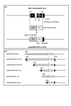
Figure 1
Figure 1
Construction of a myf5:GFP bacterial artificial chromosome (BAC) and deletion constructs for germ-line transmission in zebrafish. (A) Strategy for constructing a myf5 BAC clone containing the green fluorescent protein (GFP) reporter. (Top) The genomic organization of the myf5 that contains 3 exons (E1, E2, and E3) and 2 introns (I1 and I2). (Bottom) The resulting p(myf5(80K):GFP) clone contains the myf5 upstream 80-kb regions fused with the GFP reporter gene. The primers ZMFP-117F, GFP-R, Kan-F, and ZMF-1000R were used to check recombinants. (B) Deletion constructs used in this study. Plasmid pZMYP-2456E was described by Wang et al. [13]. Thick lines and crossed boxes represented plasmid vectors and myf5 promoters, respectively. Numbers above the boxes indicate the nucleotide positions relative to the transcription start site of zebrafish myf5. GFP, green fluorescent protein; ITR, inverted terminal repeats of adeno-associated virus; SVpA, polyadenylation signal of SV40.
Figure 2
Figure 2
Green fluorescent protein (GFP) expression in Tg(myf5(80K):GFP) transgenic embryos recapitulates endogenous myf5 expression in muscle precursors. GFP fluorescence is detected in the presomitic mesoderm of embryos by 10.5 hours postfertilization (hpf) (A), in the somites and the presomitic mesoderm in 16 hpf embryos (B). Endogenous myf5 transcripts (C,D,E) and GFP mRNA (F,G) were detectable at 7.5, 10.5, and 16 hpf. (A,D,G) Dorsal views, rostral to the left; (B,C,E,F) side views, rostral to the left, dorsal to the top. Scale bars: 100 μm in all panels.
Figure 3
Figure 3
Tg(myf5(80k):GFP) transgenic embryos express green fluorescent protein (GFP) in both slow and fast muscle fibers. (A,B) GFP expression in somites labeled with the F59 antibody. White dash lines indicate the location of head. (B) Higher magnification view of the boxed region shown in A. (C-E) Cross-section along the plane indicated by the white line in panel (B). GFP signals are observed in both fast (C, green signals) and slow muscle fibers (F, yellow signals). (A,B) Side views, rostral to the left, dorsal to the top; (C-D) dorsal to the top. 28 hpf. Scale bars: 400 μm in A; 200 μm in B; 100 μm in C-E.
Figure 4
Figure 4
Expression of green fluorescent protein (GFP) in Tg(myf5(80K):GFP) transgenics matches the dynamic pattern of endogenous myf5 expression in cranial muscles. (A-D) GFP fluorescence is apparent in pectoral fin muscle (pm), dorsal rostral muscle (drm), and hypaxial muscle (hy). (C-E) GFP fluorescence is detected in the occipital somite (os; precursors of sternohyoideus, sh) and some cranial muscles, such as the superior oblique (so) and inferior oblique (io). (F-H) Endogenous myf5 transcripts are also detected in cranial muscles, including so and io by whole-mount mRNA in situ hybridization. (A,C,F) Side views, rostral to the left, dorsal to the top; (B,D,G) dorsal views, rostral to the left; (E,H) ventral views, rostral to the left. Scale bars: 200 μm.
Figure 5
Figure 5
Green fluorescent protein (GFP) persists in cranial muscles of Tg(myf5(80k):GFP) transgenics. (A,B) GFP fluorescence is apparent in adductor hyomandibulae (ah), adductor mandibulae (am), adductor operculi (ao), constrictor hyoideus ventralis (chv), dilatator operculi (do), inferior oblique (io), lateral rectus (lr), medial rectus (mr), sternohyoideus (sh), superior oblique (so), and transverse ventralis (tv1–5) at 60 hours postfertilization (hpf). (C, D) Endogenous myf5 transcripts and (F, G) GFP mRNA are restricted to four spots by 60 hpf using whole-mount mRNA in situ hybridization. (E) At 60 hpf, myod transcripts are detected in most if not all cranial muscles. (A,C,E,F) Side views, rostral to the left, dorsal to the top; (B,D,G) ventral view, rostral to the left. Scale bars: 200 μm in all panels.
Figure 6
Figure 6
A proximal element regulates myf5 expression in the presomitic mesoderm. Green fluorescent protein (GFP) fluorescence is detected in the presomitic and somitic mesoderm of transgenic embryos harboring (A) -80 kb, Tg(myf5(80K):GFP) or (B) -10 kb, Tg(myf5(10K):GFP). Tg(myf5(10K):GFP) embryos also express GFP in the notochord. Dorsal views, rostral to the left. Scale bars: 200 μm.
Figure 7
Figure 7
The myf5 upstream region contains modules that repress expression in notochord. Green fluorescent protein (GFP) fluorescence is detected in the notochord of transgenic embryos harboring (A) -10 kb, Tg(myf5(10K):GFP), (B) -6 kb, Tg(myf5(6K):GFP), or (C-D) -3 kb, Tg(myf5(3K):GFP). Side views, rostral to the left. Scale bars: 400 μm in A; 200 μm in B, C, D.
Figure 8
Figure 8
The myf5 upstream region contains modules that regulate expression in spinal cord, bones, eyes and olfactory-pits. (A,B) Green fluorescent protein (GFP) fluorescence at 48 hours postfertilization (hpf) (A) and 72 hpf (B); the star in A indicates the location of hindbrain. (C,D) Cross-sections along the plane indicated by lines C and D in B, GFP signals are apparent in spinal cord (sc) and surface ectoderm. (E-J) GFP expression is observed in bones at 21 dpf (E and F), in bones at 60 dpf (G and H) in eyes (H and I), and olfactory pits (J). (K) The same embryo as (J) with brightfield illumination. am, adductor mandibulae; bh, basihyal; mc, Meckel's cartilage; n, notochord; op, olfactory pits; pq, palatoquadrate; sc, spinal cord. (A,B,E,H) Lateral views, rostral to the left, dorsal to the top; (F,G) ventral views, rostral to the left; (I-K) frontal views, dorsal to the top. Scare bars: 500 μm in A, E-H; 250 μm in I; 200 μm in B; 100 μm in C, D, J, K.
Figure 9
Figure 9
Multiple upstream modules regulate zebrafish myf5 expression. Thick horizontal lines, blank boxes, and solid boxes represent myf5 upstream and downstream regions, mrf4 and myf5, respectively. Numbers above crossed boxes indicate nucleotide positions relative to the transcription start site of myf5.

References

    1. Pownall ME, Gustafsson MK, Emerson CP., Jr Myogenic regulatory factors and the specification of muscle progenitors in verterbrate embryos. Annu Rev Cell Dev Biol. 2002;18:747–783. doi: 10.1146/annurev.cellbio.18.012502.105758. - DOI - PubMed
    1. Conway K, Pin C, Kiernan JA, Merrifield P. The E protein HEB is preferentially expressed in developing muscle. Differentiation. 2004;72:327–340. doi: 10.1111/j.1432-0436.2004.07207004.x. - DOI - PubMed
    1. Berkes CA, Tapscott SJ. MyoD and the transcriptional control of myogenesis. Semin Cell Dev Biol. 2005;16:585–595. doi: 10.1016/j.semcdb.2005.07.006. - DOI - PubMed
    1. Chen YH, Lee WC, Liu CF, Tsai HJ. Molecular structure, dynamic expression and promoter analysis of zebrafish (Danio rerio) myf-5 gene. Genesis. 2001;29:22–35. doi: 10.1002/1526-968X(200101)29:1<22::AID-GENE1002>3.0.CO;2-Z. - DOI - PubMed
    1. Weinberg ES, Allende ML, Kelly CS, Abdelhamid A, Murakami T, Andermann P, Doerre OG, Gruwald DJ, Riggleman B. Developmental regulation of zebrafish MyoD in wild-type, no tail and spadetail embryos. Development. 1996;122:271–280. - PubMed

Publication types

MeSH terms

Substances

LinkOut - more resources