. 2007 Jan;170(1):52-64.
doi: 10.2353/ajpath.2007.060573.

Alternative complement pathway in the pathogenesis of disease mediated by anti-neutrophil cytoplasmic autoantibodies

Affiliations

Alternative complement pathway in the pathogenesis of disease mediated by anti-neutrophil cytoplasmic autoantibodies

Hong Xiao et al. Am J Pathol. 2007 Jan.

Abstract

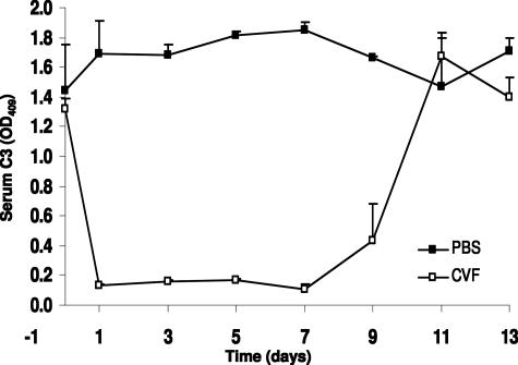
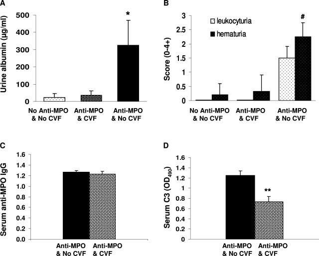
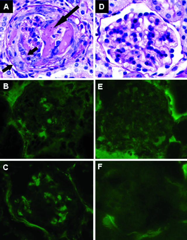
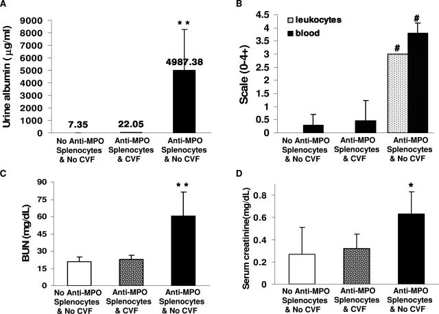
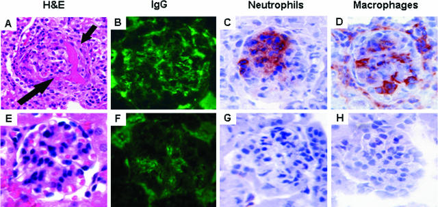
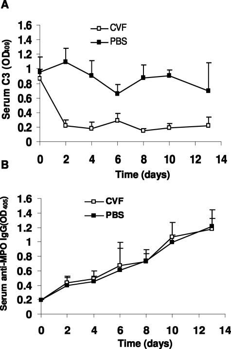
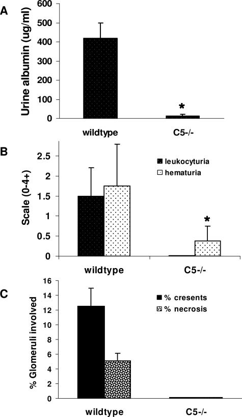
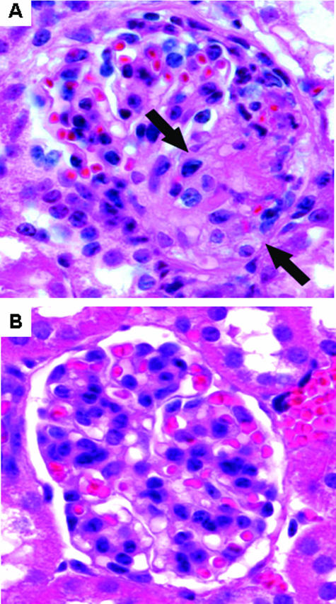
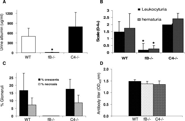
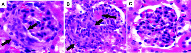
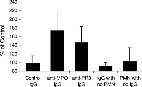
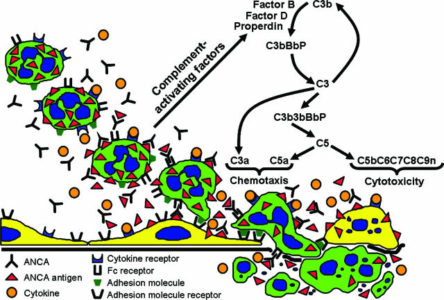
Clinical and experimental data indicate that anti-neutrophil cytoplasmic autoantibodies (ANCAs) cause glomerulonephritis and vasculitis. Here we report the first evidence that complement is an important mediator of ANCA disease. Transfer of anti-myeloperoxidase (MPO) IgG into wild-type mice or anti-MPO splenocytes into immune-deficient mice caused crescentic glomerulonephritis that could be completely blocked by complement depletion. The role of specific complement activation pathways was investigated using mice with knockout of the common pathway component C5, classic and lectin binding pathway component C4, and alternative pathway component factor B. After injection of anti-MPO IgG, C4-/- mice developed disease comparable with wild-type disease; however, C5-/- and factor B-/- mice developed no disease. To substantiate a role for complement in human ANCA disease, IgG was isolated from patients with myeloperoxidase ANCA (MPO-ANCA) or proteinase 3 ANCA (PR3-ANCA) and from controls. Incubation of MPO-ANCA or PR3-ANCA IgG with human neutrophils caused release of factors that activated complement. IgG from healthy controls did not produce this effect. The findings suggest that stimulation of neutrophils by ANCA causes release of factors that activate complement via the alternative pathway, thus initiating an inflammatory amplification loop that mediates the severe necrotizing inflammation of ANCA disease.


Figures

Figure 1
Figure 1
Profile of depletion of circulating complements by CVF. WT mice were given a single intraperitoneal injected dose of CVF (30 mg/0.3 ml PBS) or PBS (0.3 ml) and the kinetics of complement depletion were monitored by ELISA analysis of circulating C3 at indicated days after injection. Open squares represent data from CVF-treated mice (n = 6). Filled squares represent control mice that received PBS (n = 6). Bars indicate the SD.
Figure 2
Figure 2
Mice depleted of complement are resistant to anti-MPO IgG-induced experimental glomerulonephritis. WT mice were pretreated with a single dose of CVF or PBS (n = 7 for each group). Four hours later, the mice received anti-MPO IgG (50 μg/g body weight). A and B: Urine analysis showed that untreated mice and complement-depleted mice that received anti-MPO IgG had no urine abnormalities, whereas noncomplement-depleted mice that received anti-MPO IgG had albuminuria, hematuria, and leukocyturia. C and D: ELISA analysis demonstrated the same circulating anti-MPO IgG titers in noncomplement-depleted and complement-depleted mice (C); and low circulating C3 in complement-depleted mice (D). Extent of hematuria and leukocyturia is expressed as the mean on a scale of 0 (none) to 4 (severe). *P < 0.05 and #P < 0.001 versus untreated WT. **P < 0.004 compared with noncomplement-depleted mice. Bars represent the mean ± SD.
Figure 3
Figure 3
Renal tissue was examined 6 days after mice received anti-MPO IgG. WT mice that received pathogenic IgG without complement depletion developed focal glomerular necrosis (long arrow) and crescents (short arrows) (A) and low-level glomerular IgG (B) and C3 (C) deposition. In contrast, the mice depleted of complement had no glomerular lesions after the same dose of anti-MPO IgG (D) and had no IgG (E) or C3 (F) in glomeruli. A and D are stained with PAS.
Figure 4
Figure 4
Complement depletion by CVF abolishes the development of urine abnormalities, and elevation of serum blood urea nitrogen and creatinine in Rag2−/− mice 13 days after they received 5 × 10⁷ anti-MPO splenocytes (A–D). Bars represent the SD. *P < 0.05, **P < 0.01, #P < 0.001 compared with untreated Rag2−/− mice (ie, with no anti-MPO splenocytes and no CVF).
Figure 5
Figure 5
Complement depletion prevents anti-MPO splenocyte-induced glomerular necrosis and crescents. Rag2−/− mice were treated with CVF or PBS (n = 8 per group), and then, 4 hours later, were injected with 5 × 10⁷ anti-MPO splenocytes. Pathological examinations were performed at day 13 of receiving anti-MPO splenocytes. In noncomplement-depleted Rag2−/− mice, transfer of anti-MPO splenocytes induced glomerular necrosis (long arrow) and crescent formation (short arrow) (A) (H&E), moderate glomerular IgG deposition (B) (fluorescein isothiocyanate anti-IgG), and neutrophil and macrophage infiltration (C, D). Complement-depleted Rag2−/− mice that received anti-MPO splenocytes had no lesions by light microscopy (E) (H&E), low-level IgG deposition (F), and no significant increase in glomerular neutrophils or macrophages (G, H).
Figure 6
Figure 6
Circulating C3 and anti-MPO IgG levels in Rag2−/− mice during induction of NCGN. Rag2−/− mice that received pretreatment of CVF or PBS (n = 8 for each group) were injected with 5 × 10⁷ anti-MPO splenocytes. Circulating C3 and anti-MPO IgG were monitored by ELISA at different time points up to 13 days after receiving splenocytes. A: Circulating C3 levels in CVF-treated Rag2−/− mice (open squares) and control Rag2−/− mice (filled squares). B: Circulating anti-MPO IgG levels in CVF-treated Rag2−/− mice (open squares) and control Rag2−/− mice (filled squares). Bars represent the SD.
Figure 7
Figure 7
Lack of C5 blocked induction of NCGN by anti-MPO IgG. WT (n = 8) and C5−/− (n = 7) mice were injected intravenously with anti-MPO IgG. Six days after injection, urine and kidney samples were taken and examined. A and B: Urine albumin was determined by ELISA (A), and leukocyturia and hematuria were analyzed by dipsticks and expressed as the mean on a scale of 0 (none) to 4 (severe) (B). C: The extent of glomerular crescents and necrosis were expressed as the mean percentage of glomeruli with necrosis and crescents. Bars represent the SD. *P < 0.004.
Figure 8
Figure 8
Six days after injection of anti-Mpo IgG, histological examination revealed glomerular necrosis and crescents (arrows) in all WT mice (A) but no glomerular lesion in C5−/− mice that received the same dose of anti-MPO IgG (B). H&E stain.
Figure 9
Figure 9
Mice deficient in fB but not C4 were resistant to anti-MPO IgG-induced NCGN. WT (n = 8), fB−/− (n = 8), and C4−/− (n = 6) mice were injected intravenously with anti-MPO IgG and examined 6 days later. A and B: WT and C4−/− mice that received anti-MPO IgG showed albuminuria, leukocyturia, and hematuria, but fB−/− mice that received anti-MPO antibody had no urine abnormalities. Crescents and necrosis were observed in glomeruli of all WT and C4−/− mice that received anti-MPO IgG. No glomerular injury was seen in fB−/− mice injected with same amount of anti-MPO IgG. C: The extent of glomerular damage was expressed as the mean percentage of glomeruli with crescents and necrosis. D: Circulating anti-MPO antibody titers determined by ELISA were similar in WT, fB−/−, and C4−/− mice. Bars represent the SD. *P < 0.005.
Figure 10
Figure 10
A and B: Crescents and necrosis were observed in glomeruli of all WT mice (A) and all C4−/− mice (B) that received anti-MPO IgG (arrows). C: No glomerular injury was seen in fB−/− mice injected with same amount of anti-MPO IgG. H&E stain.
Figure 11
Figure 11
Incubation of TNF-α-primed normal human neutrophils with human anti-MPO IgG or anti-PR3 IgG caused release of factors that caused complement activation in normal serum as detected by generation of C3a. Normal TNF-α-primed neutrophils were first incubated with IgG and then the supernatant was reacted with normal serum and the activation of complement measured by C3a ELISA. C3a generation is expressed as a percentage of the mean of the results for control IgG. Anti-MPO IgG and anti-PR3 IgG caused C3a generation at 173.3 and 146.4%, respectively, compared with control IgG. The normal control replicate assays averaged 98.2%. The C3a generation by both anti-MPO and anti-PR3 ANCA was statistically significant compared with control (P = 0.0016). No C3a generation was caused by anti-MPO IgG, anti-PR3 IgG, or normal control IgG alone; or by TNF-α-primed neutrophils in the absence of IgG.
Figure 12
Figure 12
Diagram depicting a putative pathogenic mechanism for ANCA glomerulonephritis and vasculitis. Beginning in the top left, neutrophils are primed by cytokines to express more ANCA antigens (MPO and PR3) at the surface where they can interact with ANCA antibodies. This results in neutrophil activation both by Fc receptor engagement and Fab′2 binding. ANCA-activated neutrophils release factors (eg, properdin, proteases, oxygen radicals, and MPO) that activate the alternative complement pathway with the generation of the powerful neutrophil chemoattractant C5a and the membrane attack complex C5b-9. This complement activation amplifies neutrophil influx, neutrophil activation, and vessel damage, resulting in the aggressive necrotizing inflammation of ANCA disease.

Comment in

References

    1. Jennette JC, Xiao H, Falk RJ. The pathogenesis of vascular inflammation by antineutrophil cytoplasmic antibodies. J Am Soc Nephrol. 2006;17:1235–1242. - PubMed
    1. Bansal PJ, Tobin MC. Neonatal microscopic polyangiitis secondary to transfer of maternal myeloperoxidase-antineutrophil cytoplasmic antibody resulting in neonatal pulmonary hemorrhage and renal involvement. Ann Allergy Asthma Immunology. 2004;93:398–401. - PubMed
    1. Schlieben DJ, Korbet SM, Kimura RE, Schwartz MM, Lewis EJ. Pulmonary-renal syndrome in a newborn with placental transmission of ANCAs. Am J Kidney Dis. 2005;45:758–761. - PubMed
    1. Xiao H, Heeringa P, Hu P, Liu Z, Zhao M, Aratani Y, Maeda N, Falk RJ, Jennette JC. Antineutrophil cytoplasmic autoantibodies specific for myeloperoxidase cause glomerulonephritis and vasculitis in mice. J Clin Invest. 2002;110:955–963. - PMC - PubMed
    1. Little MA, Smyth CL, Yadav R, Ambrose L, Cook HT, Nourshargh S, Pusey CD. Anti-neutrophil cytoplasm antibodies directed against myeloperoxidase augment leukocyte-microvascular interactions in vivo. Blood. 2005;106:2050–2058. - PubMed

Publication types