. 2007 Jan;170(1):100-9.
doi: 10.2353/ajpath.2007.060806.

Aggregated myocilin induces russell bodies and causes apoptosis: implications for the pathogenesis of myocilin-caused primary open-angle glaucoma

Affiliations

Aggregated myocilin induces russell bodies and causes apoptosis: implications for the pathogenesis of myocilin-caused primary open-angle glaucoma

Gary Hin-Fai Yam et al. Am J Pathol. 2007 Jan.

Abstract

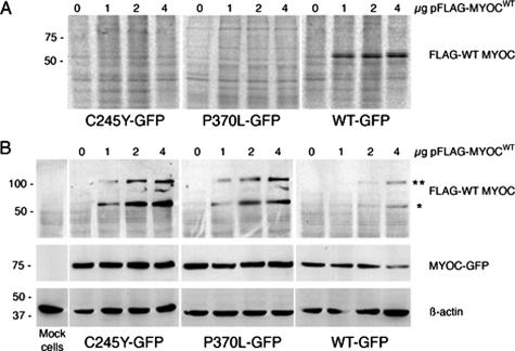
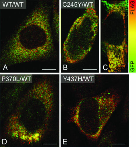
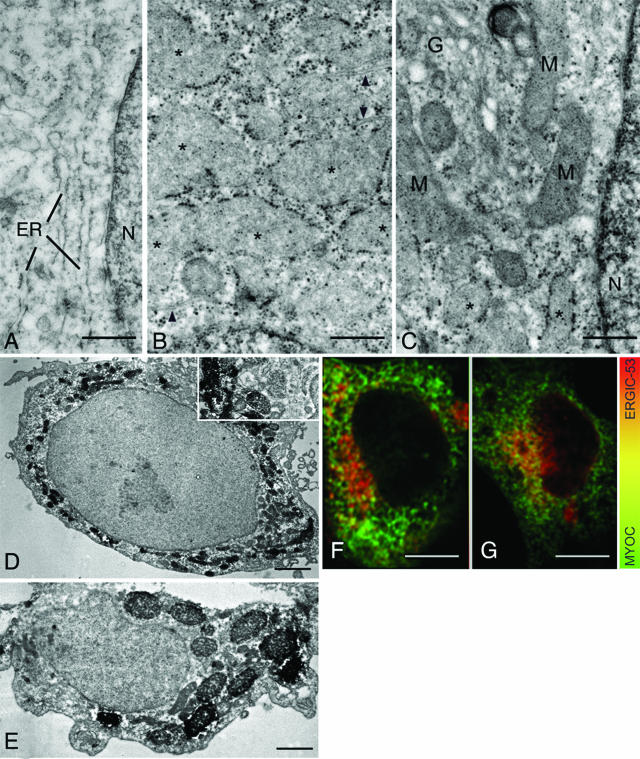
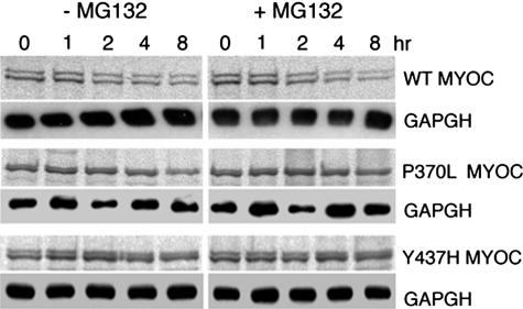
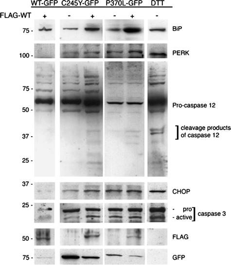
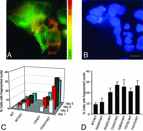
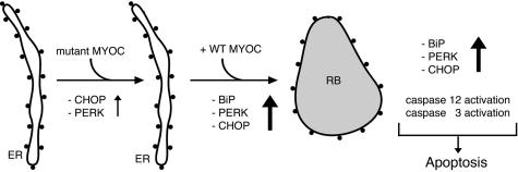
Primary open-angle glaucoma with elevated intraocular pressure is a leading cause of blindness worldwide. Mutations of myocilin are known to play a critical role in the manifestation of the disease. Misfolded mutant myocilin forms secretion-incompetent intracellular aggregates. The block of myocilin secretion was proposed to alter the extracellular matrix environment of the trabecular meshwork, with subsequent impediment of aqueous humor outflow leading to elevated intraocular pressure. However, the molecular pathogenesis of myocilin-caused glaucoma is poorly defined. In this study, we show that heteromeric complexes composed of wild-type and mutant myocilin were retained in the rough endoplasmic reticulum, aggregating to form inclusion bodies typical of Russell bodies. The presence of myocilin aggregates induced the unfolded protein response proteins BiP and phosphorylated endoplasmic reticulum-localized eukaryotic initiation factor-2alpha kinase (PERK) with the subsequent activation of caspases 12 and 3 and expression of C/EBP homologous protein (CHOP)/GADD153, leading to apoptosis. Our findings identify endoplasmic reticulum stress-induced apoptosis as a pathway to explain the reduction of trabecular meshwork cells in patients with myocilin-caused glaucoma. As a consequence, the phagocytotic capacity of the remaining trabecular meshwork cell population would be insufficient for effective cleaning of aqueous humor, constituting a major pathogenetic factor for the development of increased intraocular pressure in primary open-angle glaucoma.


Figures

Figure 1
Figure 1
Heteromeric complexes of mutant and WT MYOC are not secreted and form detergent-insoluble aggregates. A: CHO cells stably expressing either mutant (C245Y, P370L) or WT MYOC-GFP were transfected with increasing amounts of pFLAG-MYOCWT for 2 days in culture media containing 25 μCi/ml [35S]cysteine and [35S]methionine. The culture media were immunoprecipitated with anti-FLAG antibody, the samples resolved by 10% SDS-PAGE, and analyzed by phosphorimaging. B: Same material as in A. Cells were lysed in buffer containing 0.5% Triton X-100 and detergent-insoluble fractions were analyzed for FLAG, GFP, and β-actin by Western blotting. *Monomeric FLAG-WT MYOC, and **heteromeric FLAG-WT MYOC/MYOC-GFP complexes. The staining for GFP and β-actin served as loading control.
Figure 2
Figure 2
Co-expression of mutant and WT MYOC results in intracellular aggregate formation. A: Merged double-confocal immunofluorescence images of HEK293 cells expressing WT MYOC-GFP (green) and FLAG-WT MYOC (red) shows overlapping MYOC distribution as indicated by the orange-yellow color. MYOC aggregates are undetectable. B and C: C245Y- and WT MYOC-expressing cells exhibit distinct cytoplasmic aggregates composed of both mutant and WT MYOC as indicated by the orange-yellow color. Distinct cytoplasmic aggregates of varying sizes are also observed in P370L and WT MYOC-expressing (D) or Y437H-expressing and WT MYOC-expressing cells (E). Scale bars = 5 μm.
Figure 3
Figure 3
Heteromeric complexes of mutant/WT MYOC aggregate in rough ER-derived Russell bodies. A: The lumen of the rough ER in WT/WT MYOC-expressing HEK293 cells is narrow. B and C: The lumen of ribosome-covered ER in C245Y/WT MYOC-expressing HEK293 cells is greatly expanded (asterisks). Some adjacent cisternae exhibit a narrow lumen (arrowheads). The pre-Golgi intermediate, Golgi cisternal stack (G in C), and nuclear envelope appear normal in cells with dilated ER cisternae (asterisks). D and E: Immunoperoxidase electron microscopy reveals the presence of FLAG-WT MYOC in the dilated cisternae of the ER and Russell bodies. F and G: Confocal double immunofluorescence for MYOC and ERGIC-53 in C245Y/WT MYOC-expressing (E) and WT/WT MYOC-expressing cells (F). M, mitochondria; N, nucleus. Scale bars = 0.1 μm (A–C); 1 μm (D, E); 5 μm (F, G).
Figure 4
Figure 4
Turnover of WT and mutant MYOC-GFP is not influenced by proteasome inhibition. Autoradiogram of metabolically labeled and immunoprecipitated WT, P370L, and Y437H MYOC chased in the presence of 10 μmol/L MG132. Identical results were obtained for HTM cells (shown here) as well as HEK293 and CHO cells (not shown). Western blotting of GAPDH in soluble cell lysates served as loading control.
Figure 5
Figure 5
Heteromeric mutant/WT MYOC aggregates up-regulate unfolded protein response components. RIPA soluble lysates of WT/WT, C245Y/WT, and P370L/WT MYOC-expressing HEK293 cells were subjected to Western blotting for BiP, PERK, caspase 12, CHOP/GADD153, and caspase 3. The staining for FLAG and GFP demonstrates WT and mutant MYOC expression in these cells. Dithiothreitol-treated mock-transfected cells served as positive control for ER stress induction.
Figure 6
Figure 6
Heteromeric mutant/WT MYOC aggregates induce apoptosis. A: HEK293 cells stably expressing only C245Y MYOC-GFP appear in green whereas those co-expressing the mutant and the FLAG-WT MYOC can be identified by their orange-yellow color. B: The some cluster of cells as shown in A but stained by Hoechst 332518 DNA stain. The C245Y MYOC-GFP and FLAG-WT MYOC co-expressing cells exhibit fragmented nuclei C: Apoptosis rate was highest in C245Y/WT-expressing cells when compared with WT, WT/WT, or C245Y MYOC-expressing cells and increased with time after transfection. D: Apoptosis rate of different mutant/WT MYOC-expressing cells at day 5 after pFLAG-MYOCWT transfection.
Figure 7
Figure 7
Summary of the proposed mechanism of mutant MYOC-caused apoptotic cell death. In cells expressing the ER-retained mutant MYOC only, PERK and CHOP are weakly induced. However, in cells co-expressing Russell body (RB)-forming, detergent-insoluble heteromeric aggregates of mutant and WT MYOC, BiP, PERK, and CHOP are strongly induced. In addition, activation of caspases 12 and 3 occurs. Eventually, the induction of unfolded protein response components and apoptosis mediators results in apoptotic cell death.

References

    1. Shields MB, Ritch R, Krupin T. Classification of the Glaucoma. Ritch R, Shields MB, Krupin T, editors. Philadelphia: Mosby,; 1996:pp 717–725.
    1. Quigley HA. Number of people with glaucoma worldwide. Br J Ophthalmol. 1996;80:389–393. - PMC - PubMed
    1. Tamm ER. Myocilin and glaucoma: facts and ideas. Prog Retin Eye Res. 2002;21:395–428. - PubMed
    1. Tan JC, Peters DM, Kaufman PL. Recent developments in understanding the pathophysiology of elevated intraocular pressure. Curr Opin Ophthalmol. 2006;17:168–174. - PubMed
    1. Grierson I, Hogg P. The proliferative and migratory activities of trabecular meshwork cells. Prog Retin Eye Res. 1995;15:33–67.

Publication types

MeSH terms

Substances